
Журнал «Здоровье ребенка» 7 (50) 2013
Вернуться к номеру
A case report on mallory-weiss esophageal tear in a child
Авторы: Prokhorov Ye.V., Belskaya E.А., Matsynina М.А., Poshekhonov А.S.
Рубрики: Педиатрия/Неонатология
Версия для печати
The article presents a clinical case of Mallory-Weiss esophageal tear in a 5-year-old child.
Mallory-Weiss esophageal tear, children.
Mallory-Weiss esophageal tear is one of the less common reasons for upper gastrointestinal bleeding [1]. In 1929 the American pathologist G. Kenneth Mallory (1900 -1986) and the American doctor Soma Weiss (1898 - 1942) were the first to describe a patient with gastrointestinal bleeding as a result of linear tears of mucous coat, in particular, in the cardioesophageal area. Since then such bleeding has been viewed as Mallory-Weiss esophageal tear [2]. According to modern views, the tear is regarded as a linear fissure of the mucous coat of the esophagus or stomach accompanied by bleeding due to incessant vomiting or other factors increasing the abdominal and gastric pressure [3]. Based on the data obtained by I.V. Mayev and А.А. Samsonov in their pediatric practical work, Mallory-Weiss esophageal tear accounts for 5% of all the cases of bleeding [4].
A.E. Bharucha, C.J. Gostout and R.K. Balm [5] view Mallory-Weiss esophageal tear as a stomach trauma, preconditioned by an abrupt increase in the gastric pressure of the hydrodynamic shock type. Alongside vomiting attacks, abdominal pressure can rise in case of incessant vomiting, bouts of coughing, asthmatic fits, a blunt abdominal trauma, defecation strain, weightlifting, an epileptic seizure etc. [2,5].
The development of a real pathology in children is believed to be preconditioned by a decrease in the resistance of the upper gastrointestinal mucous coat, which can be predetermined by overeating, medication (aspirin, corticosteroids), hiatal hernia, respiratory and cardiovascular disorders, chronic esophagitis etc. [3].
Over the past 5 years Donetsk Regional Children’s Clinical Hospital has registered 3 cases of the tear in question. Due to the relative rarity of the pathology and the insufficient awareness of the syndrome on the part of the medical staff, we would like to present the results of our observation of Mallory-Weiss esophageal tear.
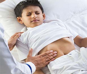
The boy A., aged 5, was admitted to the Casualty Department of Donetsk Regional Children’s Clinical Hospital (Medical History #1836) with complaints about coughing, a running nose, fever of 38,0 С0, repeated haematemesis, tarry stool.
The boy had fallen ill two days before, when his temperature rose to 38,5 С0, coughing appeared. The child was given antiviral, expectorant and antipyretic medicines. At the peak of a coughing fit the boy started vomiting with scarlet blood mixed in. The child was taken to hospital, where he underwent urgent esophagogastroduodenoscopy. Research Protocol: the insertion of the apparatus into the esophageal lumen revealed liquid blood. Washing the walls of the esophagus with physiological solution revealed no defects of the mucous coat. The stomach contained a large quantity of liquid red blood, which was aspirated. On the bottom of the stomach there were two linear defects up to 5 cm, which constantly leaked blood. The mucous coat and the antrum of the stomach did not reveal any visible defects. The duodenal bulb had the right shape, no defects of the mucous coat were detected. Features of Mallory-Weiss esophageal tear present, bleeding.
At the point of admission, the condition of the patient was critical, which was predetermined by gastric bleeding and features of anaemic syndrome connected with the acute blood loss. Breathing rate – 25 per minute, heart rate – 95 per minute, blood pressure - 85/50 mm of mercury. Clear-cut catarrhal signs in the epipharynx. Fits of cough. Profuse vomiting with scarlet blood mixed in. Skin cover and mucous coats were pale. During auscultation – harsh breathing, dry and sporadical medium moist rales on both sides. Moderately muffled and rapid heart tones, systolic murmur at I and V points. While palpated, the abdomen was soft, painful sensations present in the paraumblical area. The liver was located close to the edge of the arch of ribs, spleen was not palpable.
General blood test: red blood cells – 3,5 grams per liter, hemoglobin – 90 grams per liter, white blood cells – 11,6 grams per liter. Hematocrit – chest X-ray revealed features of bronchitis.
Clinical diagnosis: Mallory-Weiss esophageal tear, hemorrhagic anemia.
Secondary diagnosis: Acute bronchitis.
Due to the seriousness of his condition, the child was taken to the Resuscitation Unit, where he underwent a mucolytic, haemostatic (Vicasol, aminocapronic acid, Dicynon, quarantine fresh-frozen plasma), substitutive (packed red blood cells) therapy. On the fifth day the boy was transferred to the specialized somatic unit, where the therapy was continued with antacids and regenerative medicines.
After 10 days an esophagogastroduodenoscopy confirmed a normal endoscopic state of the esophageal, gastric and duodenal mucous coats. The child was discharged in the satisfactory condition.
In this case the complication (Mallory-Weiss esophageal tear) can be regarded as a result of an increase in the abdominal pressure caused by acute bronchitis as well as the damaging effect of an antipyretic medicine on the mucous coat, resulting in the tear of the mucous coat of the gastric fundus and bleeding of the damaged vessels.