
Журнал «Здоровье ребенка» 1 (52) 2014
Вернуться к номеру
Role of adrenaline in progression of arterial hypertension in children and adolescents
Авторы: I.B. Zyukova, N.N. Kaladze - Crimea State Medical University named after S.I. Georgievsky, Simferopol, Ukraine
Рубрики: Педиатрия/Неонатология
Разделы: Клинические исследования
Версия для печати
hypertension, adrenaline, children, the pressure, the adrenal glands, the sympathetic-adrenal system
As early as in 1563 Bartolomeo Eustachius, the Great anatomist of the Renaissance, established that suprarenal glands are self-depended organs, Ecker described a constitution of the suprarenal glands in fulsome details after 300 years, and Thomas Addison represented about symptoms and pathological findings, connected with the suprarenal glands destruction.
The suprarenal glands (grandulae suprarenales) are paired endocrine glands, which have a form of triangular formations and located on the upper sections of kidneys. Each suprarenal gland in mammals consists of two dissimilar parts anatomically related to each other. Although mammalian and human medullary and cortical substances are anatomically combined in one organ, functionally they are separated. There are three zones in the cortical substance: zona glomerulosa, sited outside under the adrenal capsule, zona fasciculate, which is in the middle, and reticular zone, which borderlines with the medullary substance. The barriers between the zones are arbitrary and variable. The cortical substance provides an admission of steroid hormones (glucocorticoids, mineralocorticoids, androgenic steroids with estrogenic properties) into the bloodstream.
Ental, or medullar, a part of the gland has another nature. It arises from the sympathetic ganglion and remains connected to sympathetic nerves. The adrenal medulla functions as an endocrine gland. It consists of a group of polyhedral cells containing chromaffin granules. The groups of cells are separated by blood sinuses, flowing into the central vein. The adrenal medulla is provided with ample blood supply. Within one minute, the blood, exceeding its weight in 6–7 times is delivered to the adrenal medulla. Catecholamines (adrenaline and noradrenaline) are the hormones of medulla.
In 1856 it was discovered that the adrenal medulla is discolored into emerald green with a ferrous chloride. Lesser, but similar staining, the blood, flowing from the adrenal glands, gave. It was shown, that there is a substance in adrenal glands, flowing to the blood.
In 1894 George Oliver and Edward Schaefer demonstrated vasoconstrictor and pressor effect of extract of the adrenal glands. In 1897, John Abel extracted pure adrenaline from sheep’s adrenal glands, which could quickly increase blood pressure and heart rate, improve a clear airway. Abel published the results of his experiments, and named the resulting material "epinephrine". Following Abel in 1900 Jokić Takamine developed a technology of the production of the active substance of the adrenal medulla. He described the chemical formula of the substance, gave it a name "adrenaline" and patented his invention.
Catecholamines are neurotransmitters, which mediate the function of the CNS and the sympathetic nervous system, taking the main part in the regulation of the cardiovascular system. Tyrosine, which through a series of compounds is converted into adrenaline, is a starting material for the formation of the catecholamines.
The catecholamines are settled in granules in chromaffin cells, which serve as a reservoir, a place of their biosynthesis and release. In addition to the catecholamines, the granules contain lipids, nucleotides (ATP), proteins, Ca2 + and Mg2 +. 80 % of adrenalin and 20 % of noradrenaline are contained in the granules of the adrenal medulla. A catecholamine secretion is realized by exocytosis; with that, a content of the granules “is poured” into the extracellular space. The granules have the following specific functions: they absorb a dopamine from a cytosol of the cell and convert it to noradrenaline; they are a locus of "warehousing" of adrenaline and noradrenaline; they safeguard adrenaline and noradrenaline from the effect of monoamine oxidase and destruction, and in responsiveness to nerve stimulation release the catecholamines in the blood.
In the endings of sympathetic nerve fibers, the granules, containing only norepinephrine, are revealed. The similar granules were discovered in ganglia of the sympathetic nervous system. Noradrenaline was found in the cerebrum and spinal medulla, but the maximal concentration - in the hypothalamus. The content of adrenaline in these areas is imperceptible. About 80 % of the noradrenaline contained in these spheres is localized in synaptosomes and nerve endings. It is important to note, that about 50 % of the catecholamines contained in the hypothalamus and other basal ganglia of the cerebrum is accounted for by dopamine. Release of the catecholamines from both the adrenal medulla and the sympathetic nerve endings is influenced by physiological stimulants such as stress, physical and mental stress, increased insulin levels, hypoglycemia, hypotension, etc. Release of the catecholamines occurs involving Ca2+ ions, which invade the cell or the endings of the sympathetic nervous system. The catecholamines, percolated to the blood, attain the peripheral tissues, where they are accumulated and metabolized directly proportional to the sympathetic innervation of the tissues.
Inactivation of the catecholamines occurs involving two enzyme systems of catechol-O-methyltransferase and monoamine oxidase (MAO). COMT is an intracellular enzyme that is localized in cytoplasm. It is believed that about 50 % of COMT is contained in synaptosomes of the central and peripheral nervous systems, and the rest (50–55 %) are contained in other organs – liver, kidney, intestine, spleen, salivary glands, aorta, uterus, adipose tissue, red blood cells.
Adrenaline is the main hormone (on the quantity and activity) of the adrenal medulla. The sympathetic nervous system and the adrenal medulla, form one functional sympathoadrenal system. The main role of this system is a mobilization of the body during stress (providing reaction "Fight and flight").
Effects of the adrenaline, which are realized through the membrane α-and β-adrenergic receptors, include:
- Increasing the intensity of metabolism and oxygen consumption of the cells.
- Enhancement of glycogenolysis and lipolysis, leading to increasing of concentration of glucose and fatty acids in the blood.
- Elevation of the arterial tension.
- Bronchiectasis.
- Inhibitory action on the motility and secretion of the gastrointestinal tract.
- Mydriasis.
- Increasing in the excitability of the central nervous system.
The regulation of the secretion of catecholamines in the adrenal glands is carried out by sympathetic nervous system. The specific characteristic is an absence of autonomic ganglia, which perform the role of self adrenal medulla. Thus, the secretion of the adrenaline is increased with an activation of the sympathetic nervous system (i.e., stress), highlighting the functional unity of the sympathetic-adrenal system.
The chronic activation of the sympathetic-adrenal system is a characteristic of patients with hypertension, coronary heart disease, atherosclerosis, and a high level of catecholamines is a risk factor for cardiovascular events and survival factor for them (Vizir V.A. 2001, Sirenko YU.M.2002, Karpov YU.A. 2005, Tarasenko O.O., 2009).
Taking into account, that the posterior hypothalamus is activated under the stress, which lead to increasing a tone of the sympathetic-adrenal system and extra release of norepinephrine from sympathetic nerve endings, and from the adrenal medulla – adrenaline, we investigated the level of adrenaline in children with hypertension and healthy children.
Materials and methods are:
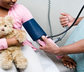
The study included 132 children with arterial hypertension (AH), aged 12–16 years (middle age is 13,87 ± 1,46). The control group (CG) consisted of 20 healthy children. Patients with the recurrent AH were not included in the study. The children were on the recovery in the “Yubileynyy” sanatorium, Evpatoria, and in the cardiorheumatological department CRU "CTH" (Children's Teaching Hospital), Simferopol. All the patients were divided according to age, sex, diagnosis. In accordance with the classification of AH, the children were divided into 2 groups at the moment of examination: labile hypertension (LH) – 71 (54 %), sustained hypertension (SH) - 61 (46 %). Boys (87 (66 %) of children) were dominated over girls (45 (34 %)) in the study group. All children underwent general clinical and laboratory studies. A fasting blood sample was being drawn from 7.30 to 8.00 am. , the serum was stored at –20 C. Determining the level of the adrenaline in the blood serum was performed using enzyme immunoassay by dint of «Adrenaline ELISA EIA» test system.
There are such results and discussion as:
Studying the level of the adrenaline in the blood serum of children with AH, we discovered its significant increase up to 5,31 ± 0,17 nmol/l (CG – 2,22 ± 0,13 nmol/l, p < 0,001) and the highest indices of the adrenaline were noted in patients with SH – 5,41 ± 0,23 nmol/l (p < 0,001); in children with LH the level of the adrenaline was also significantly higher (p < 0,001) compared to the CG – 5,02 ± 0,18 nmol/l (fig. 1).
/53/53_fig_1.jpg)
Studying the level of the adrenaline depending on sex and form of the AH the following was discovered: both the boys and the girls had significantly (p < 0,001) increase of the adrenaline in the blood serum in comparison with the indicators of CG, in a greater or lesser degree, but not significantly prominent in the group of the boys (table 1). The highest levels of the adrenaline were found in the group of boys with SH – 5,46 ± 0,27 nmol/l, which were higher in 2.3 times compared to the CG of the boys (p < 0.001). In the group of girls with LH the lowest rates of the adrenaline were found – 4,87 ± 0,36 nmol/l, but they also were higher in 2.3 times than the rates of CG of the girls.
Table 1
/53/53_fig_2.jpg)
Studying the level of the adrenaline, depending on the duration of the disease (tab. 2), following changes were identified: with a duration of the disease less than one year - 6,08 ± 0,31 nmol/l (p < 0.001), the highest rates of the adrenaline were found in children with SH. This is indicates a significant activation of the sympathetic-adrenal system, related to the development of adaptive – mechanisms to ensure homeostasis in conditions of an acute stress. With the persistent chronic stress, it leads to a stabilization of the sympathetic activity, increasing of vascular tone and myocardial injury. With the duration of the disease less than one year – 4,43 ± 0,33 nmol/l, the lowest rates of the adrenaline were found in children with LH. With the increasing of the duration of the disease, there is a growing number of a concentration level of the adrenaline in the blood serum of the children with LH. This may indicate a voltage rise and consolidation of adaptive mechanisms of the sympathetic activity with time. With the increasing of the duration of the disease more than 1 year the level of the adrenaline in children with SH decreased up to 5,51 ± 0,31 nmol/l, and in children with LH increased up to 5,33 ± 0,39 nmol/l, i.g. equalize. This is indicative of the exhaustion of functional reserves of depressant regulatory systems, which were observed with the progression of hypertension.
Table 2
/53/53_fig_3.jpg)
In consideration of the foregoing, it may be concluded that the activation of the sympathoadrenal system in the children with the arterial hypertension is defined. This is manifested in increasing of the secretion of the adrenaline. The highest concentration of the adrenaline is determined in male persons in the group of children with stable arterial hypertension and with the duration of the disease less than 1 year.