Вступ
Контрактури — це втрата рухомості суглобів, що викликана структурними змінами некісткової тканини — м’язів, зв’язок та сухожиль. Вони розвиваються, коли ці зазвичай еластичні тканини замінюються нееластичними. Все це призводить до вкорочення та твердіння цих тканин та викликає жорсткість, деформації суглобів та втрату рухомості суглобів, іноді повну.
Етіологія
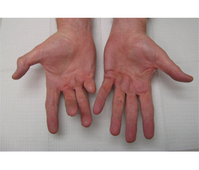
Частіше контрактури є наслідком іммобілізації, але можуть бути викликані вродженими станами (м’язовою дистрофією Дюшена, церебральним паралічем та ін.), м’язовим дисбалансом, артритами, гетеротопічною осифікацією, опіками, місцевими травмами, можуть бути наслідками тотального ендопротезування. Контрактури також можливі як наслідки впливу вібрації (контрактури Дюпюїтрена) [5, 9, 11, 13].
Епідеміологія та фактори ризику
У даний час епідеміологічна інформація в основному зосереджується на визначені суглобової конт-рактури, тому епідеміологія в цілому обмежена. До того ж розбіжності діагностичних критеріїв призводять до широкого діапазону цифр, що характеризують поширеність контрактур [3, 6].
Контрактури суглобів досить поширені. Так, за даними літератури, серед людей похилого віку вони спостерігаються у 15–70 % [7, 22], при черепно-мозковій травмі ризик розвитку контрактур становить від 16 до 80 % [17], при інсульті — біля 60 %, у пацієнтів із травмами спинного мозку можуть розвитися контрактури будь-якого суглоба в 11–50 % випадків [16]. Опікові контрактури, за даними спеціалізованих опікових відділень, становлять до 94 % [21].
Загальними факторами ризику розвитку контрактур є рухові дисфункції або тривале обмеження рухової активності, гіпоксичні ішемічні ушкодження, травми спинного мозку та похилий вік [23].
Патанатомія та фізіологія
Зміни м’яких тканин, які викликають контрактури, починаються практично одночасно з настанням нерухомості. Синтез білка у м’язових тканинах знижується через 6 годин після іммобілізації суглоба, вкорочення м’язових волокон виникає упродовж 24 годин, через 48 годин з’являється збільшена інфільтрація перимізію колагеном. Тривала нерухомість також викликає пластичні перебудови в нервових центрах, що ще більше знижує здатність залучати рухові одиниці, а це посилює вихідний парез [8, 19].
У моделях на тваринах було визначено, що 24 години ненавантаження кінцівки викликали скорочення довжини м’язових волокон на 60 % [12, 14].
Прогресування захворювання, стадії, клінічні прояви захворювання
Більшість контрактур починаються з ушкоджень тіла. Наприклад, реакція організму на болючий суглоб полягає в обмеженості його рухомості, що наражає на ризик розвитку контрактур. Неврологічні травми, що підвищують м’язовий тонус або слабкість, викликають дисбаланс м’язів, що призводить до тонічного скорочення. Отже, багатьом контрактурам суглобів передує спастичність. За відсутності лікування спастичний суглоб знерухомлюється та розвивається контрактура.
Контрактури також можуть розвиватися через невикористання кінцівки без будь-якої травми, наприклад при віковій втраті м’язової маси (саркопенія).
До специфічних вторинних чи супутніх станів та ускладнень можна віднести спастичність, гетерогенну осифікацію, дегенеративні захворювання суглобів, переломи, вивихи та інше. Деякі контрактури призводять до неправильного розташування тіла в ліжку чи інвалідному кріслі, що ще більше ускладнює стан та призводить до порочного кола, яке збільшує інвалідність.
Контрактури сильно впливають на життя людини. Ускладнення можуть викликати обмеження при проведенні гігієнічних процедур в активних людей, приводити до втрати незалежності та до постійної нерухомості.
Лікування пацієнтів із контрактурами суглобів
Анамнез повинен включати етіологію контрактури, її перебіг та вплив на функціональні можливості людини. Це — біль, обмеження пересування, повсякденної діяльності та гігієни.
При фізичному обстеженні в ідеалі у пацієнта не повинно бути болю, що може заважати повноцінному обстеженню. Огляд хворого полягає в оцінюванні розміру суглоба, симетричності в спокої порівняно з протилежною кінцівкою. Вимірюється обсяг рухів на обох кінцівках. Проводиться огляд на можливі ушкодження шкіри, що можуть бути причиною контрактур.
Але найважливішим діагностичним критерієм є пасивний діапазон рухів (ПДР, в іноземній літературі PROM — passive range of motion). Треба відзначити будь-який м’язовий дисбаланс або слабкість, підвищення тонусу чи його відсутність. Такий огляд дозволить відокремити дійсну контрактуру від контрактури, що викликана м’язовою спастикою, яка передбачає інший підхід до лікування.
Треба оцінювати активний діапазон рухів, а також силу м’язів, оскільки це може відіграти роль у визначенні причин виникнення контрактур та виборі лікування.
Методи візуалізації
Радіологічні дослідження можуть допомогти виявити патології, що ускладнюються, наприклад, кістковими деформаціями, гетеротопічною осифікацією, переломами, формуванням анкілозів. Магнітно-резонансна томографія та ультразвукова діагностика можуть використовуватися для візуалізації структур м’яких тканин з метою виявлення фіброзних змін.
Лабораторні дослідження
Діагноз «контрактура» встановлюється на основі клінічного дослідження. Немає маркерів крові або лабораторних досліджень, які б сприяли діагностиці контрактур. Але лабораторні маркери, такі як лужна фосфатаза, швидкість осідання еритроцитів та ін., можуть використовуватися для визначення таких станів, як гетеротопічна осифікація, міопатия тощо.
Додаткові інструменти оцінки
Немає перевірених інструментів оцінки контрактур, але є шкали для оцінки спастичності. Найбільш відомі з них шкала Ашворта (MAS), шкала Тардьє [15, 2] та LASIS [1].
Загальний підхід до контрактур: основні принципи
Найкраще лікування полягає в гальмуванні утворення контрактури чи запобіганні йому. Клінічно це полягає у здійсненні ПДР та щоденних фізичних вправ на розтягування. Але деякі дослідження, основані на фактичних даних, поставили ці твердження під сумнів. В оглядових статтях Кокрейна було висунуто твердження, що ПДР неефективні для профілактики та лікування контрактур [18], а розтягування не запобігає контрактурам суглобів у людей з неврологічними захворюваннями і не лікує їх [10]. Але за думкою багатьох дослідників, достатня тривалість фізичних вправ у сукупності з іншими лікувальними процедурами здатні збільшити обсяг рухів у контрактованому суглобі, і ці методи й сьогодні залишаються основними в лікуванні контрактур.
Іншим, часто основним підходом у лікуванні конт-рактур є збільшення сили м’язів, особливо антагоністів скороченого м’яза, якщо ці методи можна використати. Додаткові заходи включають усунення чи зменшення спастичності та набряків за допомогою масажу, компресійної білизни тощо.
A.J. Skalsky, C.M. McDonald (2012) [20] сформулювали основні принципи профілактики та лікування контрактур кінцівок при нервово-м’язових захворюваннях та рекомендації щодо запобігання їм:
1. Профілактика контрактур вимагає ранньої діагностики та ініціації ПДР та накладання шин.
2. При деяких нервово-м’язових захворюваннях контрактури неминучі.
3. Розширені контрактури стають фіксованими, можуть погано піддаватися консервативному лікуванню та вимагати хірургічного втручання.
4. Контроль контрактур нижніх кінцівок важливий для мінімізації їх несприятливого впливу на самостійне пересування.
5. Статичний стан як верхніх, так і нижніх кінцівок — важлива причина утворення контрактури.
6. Легкі контрактури верхніх кінцівок неспроможні негативно вплинути на рухову функцію.
Підхід до контрактур на різних етапах лікування
Дамо класичний підхід до контрактур на ранніх стадіях їх формування. Він полягає у тривалому безперервному розтягуванні суглоба за допомогою динамічної фіксації ортезом або жорсткою конструкцією з можливістю зміни кута фіксації. Процедура полягає в накладанні фіксації на максимально розігнутий суглоб, через декілька діб процедура повторюється, а суглоб розгинають на можливо більший кут. Розтягування можна комбінувати з терапевтичними процедурами нагріву, що покращує еластичність тканин.
При контрактурах, які суттєво впливають на функцію суглоба і не зменшилися після консервативних процедур, розглядають хірургічні методи лікування. До них відносяться визволення уражених м’язів, тенотомія або подовження сухожиль, визволення капсули суглоба, а в деяких випадках і повна заміна суглоба. Але хірургія часто призводить до формування вторинних контрактур через утворення рубців, ушкодження структур тканин. В останні роки все частіше застосовують малоінвазивну хірургію для запобігання великим ушкодженням. Хворі після оперативного лікування контрактур потребують більш інтенсивної реабілітації [4].
Контрактури верхніх кінцівок нечасто потребують хірургічного лікування. Воно проводиться, тільки якщо обмеження ПДР заважає проведенню гігієнічних процедур, викликає біль. Альтернативою може бути застосування ферменту колагенази для ін’єкцій, отриманого з бактерій Clostridium histolyticum (колагеназа клостридій гістолітикум) і схваленого FDA для лікування контрактури Дюпюїтрена і як альтернатива хірургічному втручанню. Клінічні наслідки при ін’єкціях колагенази аналогічні хірургічному лікуванню, але мають менше ускладнень і легше переносяться пацієнтами. Обмеженням для ін’єкцій колагенази є те, що вони зазвичай робляться в одне місце, вимагають щомісячного проведення та пов’язані з ризиком автоімунної реакції.
Лікування контрактур нижніх кінцівок полягає насамперед в їх профілактиці: регулярному стоянні та/або ходьбі; пасивному розтягуванні м’язів та суглобів; позиціюванні для розгинання та протидії згинанню; шинуванні. Головне завдання в профілактиці та реабілітації контрактур нижніх кінцівок полягає в навчанні правильному положенню кінцівки при стоянні та ходьбі. Показання для хірургічного втручання аналогічні викладеним вище: контрактура не може бути усунена фізіотерапевтичними методами та її тяжкість заважає повноцінному функціонуванню.
Висновки
В огляді ми розглянули основні причини формування контрактур суглобів та основні підходи до їх профілактики та лікування. Треба відзначити, що на сьогодні ще широко застосовуються старі, в основному пасивні методи консервативного лікування. Сучасні методи, такі як радіочастотні, лазерні, біоактивні методи, ін’єкції, часто використовуються обмежено і ще не мають загального визнання практиками.
Конфлікт інтересів. Автори заявляють про відсутність конфлікту інтересів та власної фінансової зацікавленості при підготовці даної статті.
Отримано/Received 04.01.2022
Рецензовано/Revised 10.01.2022
Прийнято до друку/Accepted 14.01.2022
Список литературы
1. Коваленко А.П., Камаева О.В., Полещук Ю.Р., Ковлен Д.В. Шкалы и тесты в реабилитации и лечении пациентов со спастичностью верхней конечности. Журнал неврологии и психиатрии им. С.С. Корсакова. 2020. 120(4). 107-114. doi:10.17116/jnevro2020120041107.
2. Коваленко А.П., Мисиков В.К., Искра Д.А., Кошкарев М.А., Синельников К.А. Шкала Тардье в диагностике спастичности. Журнал неврологии и психиатрии им. С.С. Корсакова. 2019. 119 (9). 83-90. Doi: 10.17116/jnevro201911909183.
3. Araco A., Caruso R., Araco F., Overton J., Gravante G. Capsular contractures: a systematic review. Plast. Reconstr. Surg. 2009. 124(6). 1808-1819. doi: 10.1097/PRS.0b013e3181bf7f26.
4. Degreef I. Collagenase Treatment in Dupuytren Contractures: A Review of the Current State Versus Future Needs. Rheumatol. Ther. 2016. 3(1). 43-51.
5. Descatha A., Jauffret P., Chastang J.F., Roquelaure Y., Leclerc A. Should we consider Dupuytren’s contracture as work-related? A review and meta-analysis of an old debate. BMC Musculoskelet Disord. 2011. 12. 96. Doi: 10.1186/1471-2474-12-96.
6. Fergusson D., Hutton B., Drodge A. The epidemiology of major joint contractures: a systematic review of the literature. Clin. Orthop. Relat. Res. 2007. 456. 22-9. doi: 10.1097/BLO.0b013e3180308456.
7. Gnass I., Bartoszek G., Thiesemann R., Meyer G. Joint contractures in older age. A systematic literature review [German]. Z. Gerontol. Geriatr. 2010. 43(3). 147-157.
8. Gracies J.M. Pathophysiology of spastic paresis. Paresis and soft tissue changes. Muscle Nerve. 2005. 31(5). 535-551.
9. Gudmundsson K.G., Arngrimsson R., Sigfusson N., Bjornsson A., Jonsson T. Epidemiology of Dupuytren’s disease: clinical, serological, and social assessment: The Reykjavik Study. J. Clin. Epidemiol. 2000. 53(3). 291-296. DOI: 10.1016/s0895-4356(99)00145-6].
10. Katalinic O.M., Harvey L.A., Herbert R.D., Moseley A.M., Lannin N.A., Schurr K. Stretch for the treatment and prevention of contractures. Cochrane Database Syst. Rev. 2010. 8(9). CD007455.
11. Lanting R., Broekstra D.C., Werker P.M., van den Heuvel E.R. A systematic review and meta-analysis on the prevalence of Dupuytren disease in the general population of Western countries. Plast. Reconstr. Surg. 2014. 133(3). 593-603. DOI: 10.1097/01.prs.0000438455.37604.0f.
12. Laurin J., Gondin J., Dousset E., Decherchi P. Effect of tenotomy on metabosensitive afferent fibers from tibialis anterior muscle. Exp. Brain Res. 2008. 186(1). 87-92. doi: 10.1007/s00221-007-1210-y.].
13. Layton T., Nanchahal J.F. Recent advances in the understanding of Dupuytren’s disease. F1000 Res. 2019. 8. F1000 Faculty Rev-231. doi: 10.12688/f1000research.17779.1.
14. McLachlan E.M., Chua M. Rapid adjustment of sarcomere length in tenotomized muscles depends on an intact innervation. Neurosci Lett. 1983. 35(2). 127-133.
15. Patrick E., Ada L. The Tardieu Scale differentiates contracture from spasticity whereas the Ashworth Scale is confounded by it. Clin. Rehabil. 2006. 20(2). 173-182.
16. Pingel J., Bartels E.M., Nielsen J.B. New perspectives on the development of muscle contractures following central motor lesions. J. Physiol. 2017. 595(4). 1027-1038.
17. Pohl M., Mehrholz J. A new shoulder range of motion screening measurement: its reliability and application in the assessment of the prevalence of shoulder contractures in patients with impaired consciousness caused by severe brain damage. Arch. Phys. Med. Rehabil. 2005. 86(1). 98-104.
18. Prabhu R.K., Swaminathan N., Harvey L.A. Passive movements for the treatment and prevention of contractures. Cochrane Database Syst. Rev. 2013. 12. CD009331.
19. Roby-Brami A., Jarrassé N., Parry R. Impairment and Compensation in Dexterous Upper-Limb Function After Stroke. From the Direct Consequences of Pyramidal Tract Lesions to Behavioral Involvement of Both Upper-Limbs in Daily Activities. Front. Hum. Neurosci. 2021. 15. 662006. doi: 10.3389/fnhum.2021.662006.].
20. Skalsky A.J., McDonald C.M. Prevention and management of limb contractures in neuromuscular diseases. Phys. Med. Rehabil. Clin. N. Am. 2012. 23(3). 675-687. doi:10.1016/j.pmr.2012.06.009.
21. Tan J., Chen J., Zhou J., Song H., Deng H., Ao M., Luo G., Wu J. Joint contractures in severe burn patients with early rehabilitation intervention in one of the largest burn intensive care unit in China: a descriptive analysis. Burns Trauma. 2019. 7. 17.
22. Vanderwee K., Grypdonck M., De Bacquer D., Defloor T. The identification of older nursing home residents vulnerable for deterioration of grade 1 pressure ulcers. Journal of Clinical Nursing. 2009. 18(21). 3050-3058. doi: 10.1111/j.1365-2702.2009.02860.x.
23. Zafonte R., Elovic E.P., Lombard L. Acute care management of post-TBI spasticity. J. Head Trauma Rehabil. 2004. 19(2). 89-100.