
Журнал «Боль. Суставы. Позвоночник» Том 12, №3, 2022
Вернуться к номеру
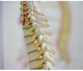
Кальцій-фосфатні кераміки в хірургії хребта: особливості регенерації і використання
Авторы: Шаповалов В.С. (1), Дєдух Н.В. (2) , Шимон М.В. (3)
(1) — Київська міська клінічна лікарня швидкої медичної допомоги, м. Київ, Україна
(2) — Інститут геронтології імені Д.Ф. Чеботарьова НАМН України, м. Київ, Україна
(3) — Ужгородський національний університет, м. Ужгород, Україна
Рубрики: Ревматология, Травматология и ортопедия
Разделы: Справочник специалиста
Версия для печати
Огляд ґрунтується на аналізі 53 джерел літератури з баз PubMed, Google, Google Scolar, Cochrane Library. Серед замінників кісткових автогенних трансплантатів у хірургії хребта керамічні біоматеріали є найбільш вивченою групою, серед них окрему нішу займають кальцій-фосфатні кераміки (КФК). Широке застосування в хірургії хребта знайшли гідроксилапатит, трикальційфосфат і біфазна кераміка з різними частинами гідроксилапатиту й трикальційфосфату. Перевагою використання КФК є їх біосумісність, остеокондуктивність, остеоіндуктивність, остеоімуномодуляція і здатність стимулювати ангіогенез — головні складові, що забезпечують регенерацію кістки. У наведеному огляді відзначені особливості регенерації в інтерфейсі «керамічний матеріал — кісткова тканина» залежно від складу, структури поверхні й кристалічності матеріалу. Позитивні результати експериментальних досліджень зумовили застосування КФК у клінічній практиці. З успіхом використовують КФК з кістковими автогенними трансплантатами при виконанні заднього й задньобічного поперекового спондилодезу, що дозволяє значно зменшити об’єм автологічної кістки. Новим напрямком є розробка неметалевих комбінованих кейджів, які використовують для виконання передньої цервікальної дискектомії і спондилодезу. До складу цих кейджів входять різні полімери в поєднанні з КФК і кістковим автогенним трансплантатом для забезпечення якісного спондилодезу, зниження стрес-шилдингу й просідання кейджу. Особливого підходу вимагає вивчення деградації і резорбції остеокластами різних КФК для керування й синхронізації процесу «резорбція — кісткоутворення». Серед питань, які потребують дослідження, необхідне подальше вивчення молекулярних механізмів остеоімуномодуляції та факторів, що стимулюють цей процес в умовах використання КФК для підвищення остеоінтеграції та остеоіндукції в керуванні репаративним остеогенезом.
The review is based on the analysis of 53 literature sources from PubMed, Google, Google Scholar, and Cochrane Library. Among the substitutes for autogenous bone grafts in spine surgery, ceramic biomaterials are the largest studied group, among which certain niche is occupied by calcium phosphate ceramics (CPCs). Hydroxylapatite, tricalcium phosphate and biphasic ceramics with all parts of hydroxylapatite and tricalcium phosphate are widely used in spine surgery. The advantage of using CPCs is their biocompatibility, osteoconductivity, osteoinductivity, osteoimmunomodulation and the ability to stimulate angiogenesis — the main components that ensure bone regeneration. In the given review, the peculiarities of regeneration in the interface “ceramic material — bone tissue” depending on the composition, surface structure, and crystallinity of the material are noted. The positive results of the experimental studies led to the use of CPCs in the clinic. CPCs with autogenous bone grafts are successfully used in posterior and posterolateral lumbar spondylodesis, which allows reducing significantly the volume of autologous bone. A new direction is the development of non-metallic combined cages, which are used to perform anterior cervical discectomy and spondylodesis. The composition of these cages includes various polymers in combination with CPCs and bone autogenous graft to ensure high-quality spondylodesis, reduce of stress-shielding and subsidence of the cage. A special approach requires the study of degradation and resorption by osteoclasts of various CPCs in order to control and synchronize the process of “resorption — bone formation”. Among the issues for the further research, the molecular mechanisms of osteoimunomodulation and factors that stimulate osseointegration and osteoinduction in the management of reparative osteogenesis should be further explored.
кальцій-фосфатні кераміки; гідроксилапатит; трикальційфосфат; модифікація керамік; регенерація; спондилодез; огляд
calcium-phosphate ceramics; hydroxylapatite; tricalcium phosphate; modification of ceramics; regeneration; spondylodesis; review
Для ознакомления с полным содержанием статьи необходимо оформить подписку на журнал.
- Eliaz N., Metoki N. Calcium Phosphate Bioceramics: A Review of Their History, Structure, Properties, Coating Technologies and Biomedical Applications. Materials (Basel). 2017. 10(4). 334. doi: 10.3390/ma10040334.
- Dong C., Klimek P., Abächerli C., De Rosa V., Krieg A.H. Percutaneous cyst aspiration with injection of two different bioresorbable bone cements in treatment of simple bone cyst. J. Child Orthop. 2020 Feb 1. 14(1). 76-84. doi: 10.1302/1863-2548.14.190155.
- Vezenkova A., Locs J. Sudoku of porous, injectable calcium phosphate cements — Path to osteoinductivity. Bioact. Mater. 2022 Jan 10. 17. 109-124. doi: 10.1016/j.bioactmat.2022.01.001.
- Ginebra M.P., Espanol M., Maazouz Y., Bergez V., Pastorino D. Bioceramics and bone healing. EFORT Open Rev. 2018 May 21. 3(5). 173-183. doi: 10.1302/2058-5241.3.170056.
- Litak J., Czyzewski W., Szymoniuk M., Pastuszak B., Litak J., Litak G., Grochowski C., Rahnama-Hezavah M., Kamieniak P. Hydroxyapatite Use in Spine Surgery-Molecular and Clinical Aspect. Materials (Basel). 2022 Apr 15. 15(8). 2906. doi: 10.3390/ma15082906.
- Ohe M., Moridaira H., Inami S., Takeuchi D., Nohara Y., Taneichi H. Pedicle screws with a thin hydroxyapatite coating for improving fixation at the bone-implant interface in the osteoporotic spine: experimental study in a porcine model. J. Neurosurg. Spine. 2018 Jun. 28(6). 679-687. doi: 10.3171/2017.10.SPINE17702.
- Lu J., Yu H., Chen C. Biological properties of calcium phosphate biomaterials for bone repair: a review. RSC Adv. 2018 Jan 9. 8(4). 2015-2033. doi: 10.1039/c7ra11278e.
- Albrektsson T., Johansson C. Osteoinduction, osteoconduction and osseointegration. Eur. Spine J. 2001 Oct. 10 Suppl 2(Suppl 2). S96-101. doi: 10.1007/s005860100282.
- Uetanabaro L.C., Claudino M., Zancan R., Zielak J.C., Garlet G.P., de Araujo M.R. Osteoconductivity of Biphasic Calcium Phosphate Ceramic Improves New Bone Formation: A Histologic, Histomorphometric, Gene Expression, and Microcomputed Tomography Study. Int. J. Oral Maxillofac. Implants. 2020 Jan/Feb. 35(1). 70-78. doi: 10.11607/jomi.7745.
- Wang W., Yeung K.W.K. Bone grafts and biomaterials substitutes for bone defect repair: A review. Bioact. Mater. 2017 Jun 7. 2(4). 224-247. doi: 10.1016/j.bioactmat.2017.05.007.
- Bohner M., Miron R.J. A proposed mechanism for material-induced heterotopic ossification. Materials Today. 2019. 22. 132-141. doi: 10.1016/j.mattod.2018.10.036.
- Bohner M., Santoni B.L.G., Döbelin N. β-tricalcium phosphate for bone substitution: Synthesis and properties. Acta Biomater. 2020 Sep 1. 113. 23-41. doi: 10.1016/j.actbio.2020.06.022.
- Gamblin A.L., Brennan M.A., Renaud A. et al. Bone tissue formation with human mesenchymal stem cells and biphasic calcium phosphate ceramics: the local implication of osteoclasts and macrophages. Biomaterials. 2014 Dec. 35(36). 9660-7. doi: 10.1016/j.biomaterials.2014.08.018.
- He Y., Peng Y., Liu L. et al. The Relationship between Osteoinduction and Vascularization: Comparing the Ectopic Bone Formation of Five Different Calcium Phosphate Biomaterials. Materials (Basel). 2022 May 10. 15(10). 3440. doi: 10.3390/ma15103440.
- Tang Z., Li X., Tan Y., Fan H., Zhang X. The material and biological characteristics of osteoinductive calcium phosphate ceramics. Regen. Biomater. 2018 Feb. 5(1). 43-59. doi: 10.1093/rb/rbx024.
- Vecbiskena L., Gross K.A., Riekstina U., Yang T.C. Crystallized nano-sized alpha-tricalcium phosphate from amorphous calcium phosphate: microstructure, cementation and cell response. Biomed. Mater. 2015 Apr 17. 10(2). 025009. doi: 10.1088/1748-6041/10/2/025009.
- Fujita R., Yokoyama A., Kawasaki T., Kohgo T. Bone augmentation osteogenesis using hydroxyapatite and beta-tricalcium phosphate blocks. J. Oral Maxillofac. Surg. 2003 Sep. 61(9). 1045-53. doi: 10.1016/s0278-2391(03)00317-3.
- Boden S.D., Schimandle J.H., Hutton W.C. An experimental lumbar intertransverse process spinal fusion model. Radiographic, histologic, and biomechanical healing characteristics. Spine (Phila Pa 1976). 1995 Feb 15. 20(4). 412-20. doi: 10.1097/00007632-199502001-00003.
- Chen Z., Klein T., Murray R.Z., et al. Osteoimmunomodulation for the development of advanced bone biomaterials. 2016 Jul-Aug. 19(6). 304-321. doi: 10.1016/j.mattod.2015.11.004.
- Xie Y., Hu C., Feng Y. et al. Osteoimmunomodulatory effects of biomaterial modification strategies on macrophage polarization and bone regeneration. Regen. Biomater. 2020 Jun. 7(3). 233-245. doi: 10.1093/rb/rbaa006.
- Arbez B., Libouban H. Behavior of macrophage and osteoblast cell lines in contact with the β-TCP biomaterial (beta-tricalcium phosphate). Morphologie. 2017 Sep. 101(334). 154-163. doi: 10.1016/j.morpho.2017.03.006.
- Duan R., Zhang Y., van Dijk L. et al. Coupling between macrophage phenotype, angiogenesis and bone formation by calcium phosphates. Mater. Sci Eng. C. Mater. Biol. Appl. 2021 Mar. 122. 111948. doi: 10.1016/j.msec.2021.111948.
- Humbert P., Brennan M.Á., Davison N. et al. Immune Modulation by Transplanted Calcium Phosphate Biomaterials and Human Mesenchymal Stromal Cells in Bone Regeneration. Front. Immunol. 2019 Apr 2. 10. 663. doi: 10.3389/fimmu.2019.00663.
- Sheikh Z., Najeeb S., Khurshid Z., Verma V., Rashid H., Glogauer M. Biodegradable Materials for Bone Repair and Tissue Engineering Applications. Materials (Basel). 2015 Aug 31. 8(9). 5744-5794. doi: 10.3390/ma8095273.
- Wang M., Chen F., Wang J. et al. Calcium phosphate altered the cytokine secretion of macrophages and influenced the homing of mesenchymal stem cells. J. Mater. Chem. B. 2018 Aug 7. 6(29). 4765-4774. doi: 10.1039/c8tb01201f.
- Wang J., Chen Y., Zhu X. et al. Effect of phase composition on protein adsorption and osteoinduction of porous calcium phosphate ceramics in mice. J. Biomed. Mater. Res. A. 2014 Dec. 102(12). 4234-43. doi: 10.1002/jbm.a.35102.
- Ogose A., Hotta T., Kawashima H. et al. Comparison of hydroxyapatite and beta tricalcium phosphate as bone substitutes after excision of bone tumors. J. Biomed. Mater. Res. B Appl. Biomater. 2005 Jan 15. 72(1). 94-101. doi: 10.1002/jbm.b.30136.
- Li X., Song T., Chen X. et al. Osteoinductivity of Porous Biphasic Calcium Phosphate Ceramic Spheres with Nanocrystalline and Their Efficacy in Guiding Bone Regeneration. ACS Appl. Mater. Interfaces. 2019 Jan 30. 11(4). 3722-3736. doi: 10.1021/acsami.8b18525.
- Chen F., Wang M., Wang J. et al. Effects of hydroxyapatite surface nano/microstructure on osteoclast formation and activity. Journal of Materials Chemistry B. 2019. 7 (47). 7574-7587. doi: 10.1039/C9TB01204D.
- Wang Z., Sakakibara T., Sudo A., Kasai Y. Porosity of β-tricalcium phosphate affects the results of lumbar posterolateral fusion. J. Spinal Disord. Tech. 2013 Apr. 26(2):E40-5. doi: 10.1097/BSD.0b013e31823db5e6.
- Kadam A., Millhouse P.W., Kepler C.K. et al. Bone substitutes and expanders in Spine Surgery: A review of their fusion efficacies. Int. J. Spine Surg. 2016 Sep 22. 10. 33. doi: 10.14444/3033.
- Van Dijk L.A., Barbieri D., Groot F.B.D. et al. Efficacy of a synthetic calcium phosphate with submicron surface topography as autograft extender in lapine posterolateral spinal fusion. J. Biomed. Mater. Res. Part B. 2019. 107B. 2080-2090. doi: 10.1002/jbm.b.34301.
- Van Dijk L.A., Duan R., Luo X. et al. Biphasic calcium phosphate with submicron surface topography in an Ovine model of instrumented posterolateral spinal fusion. JOR Spine. 2018 Nov 28. 1(4). e1039. doi: 10.1002/jsp2.1039.
- Li X., Zhou Q., Wu Y. et al. Enhanced bone regenerative properties of calcium phosphate ceramic granules in rabbit posterolateral spinal fusion through a reduction of grain size. Bioact. Mater. 2021 Oct 8. 11. 90-106. doi: 10.1016/j.bioactmat.2021.10.006.
- Sato K., Kumagai H., Funayama T. et al. Posterolateral lumbar spine fusion with unidirectional porous beta-tricalcium phosphate in a canine model. J. Artif. Organs. 2020 Dec. 23(4). 365-370. doi: 10.1007/s10047-020-01178-9.
- Dai L.Y., Jiang L.S. Single-level instrumented posterolateral fusion of lumbar spine with beta-tricalcium phosphate versus autograft: a prospective, randomized study with 3-year follow-up. Spine (Phila Pa 1976). 2008 May 20. 33(12). 1299-304. doi: 10.1097/BRS.0b013e3181732a8e.
- Yamada T., Yoshii T., Sotome S. et al. Hybrid grafting using bone marrow aspirate combined with porous β-tricalcium phosphate and trephine bone for lumbar posterolateral spinal fusion: a prospective, comparative study versus local bone grafting. Spine (Phila Pa 1976). 2012 Feb 1. 37(3). E174-179. doi: 10.1097/BRS.0b013e3182269d64.
- Epstein N.E. Beta tricalcium phosphate: observation of use in 100 posterolateral lumbar instrumented fusions. Spine J. 2009. 9(8). 630-638. doi:10.1016/j.spinee.2009.04.007.68.
- Kong S., Park J.H., Roh S.W. A prospective comparative study of radiological outcomes after instrumented posterolateral fusion mass using autologous local bone or a mixture of beta-TCP and autologous local bone in the same patient. Acta Neurochir. (Wien). 2013. 155(5). 765-770. doi:10.1007/s00701-013-1669-1.66.
- Park J.H., Choi C.G., Jeon S.R., Rhim S.C., Kim C.J., Roh S.W. Radiographic Analysis of Instrumented Posterolateral Fusion Mass Using Mixture of Local Autologous Bone and b-TCP (PolyBone®) in a Lumbar Spinal Fusion Surgery. J. Korean Neurosurg. Soc. 2011. 49(5). 267-272. doi:10.3340/jkns.2011.49.5.267.70.
- Thaler M., Lechner R., Gstöttner M., Kobel C., Bach C. The use of beta-tricalcium phosphate and bone marrow aspirate as a bone graft substitute in posterior lumbar interbody fusion. Eur. Spine J. 2013. 22(5). 1173-1182. doi: 10.1007/s00586-012-2541-3.67.
- Timothy J., Wilson J., Rice E., Hall R. Nanocrystalline hydroxyapatite intervertebral cages induce fusion after anterior cervical discectomy and may be a safe alternative to PEEK or carbon fiber intervertebral cages. Br. J. Neurosurg. 2016 Dec. 30(6). 654-657. doi: 10.3109/02688697.2016.1173192.
- Zadegan S.A., Abedi A., Jazayeri S.B., Bonaki H.N., Vaccaro A.R., Rahimi-Movaghar V. Clinical Application of Ceramics in Anterior Cervical Discectomy and Fusion: A Review and Update. Global. Spine J. 2017 Jun. 7(4). 343-349. doi: 10.1177/2192568217699201.
- Hu B., Yang X., Hu Y. et al. The n-HA/PA66 Cage Versus the PEEK Cage in Anterior Cervical Fusion with Single-Level Discectomy During 7 Years of Follow-Up. World Neurosurg. 2019 Mar. 123. e678-e684. doi: 10.1016/j.wneu.2018.11.251.
- Yamagata T., Naito K., Arima H., Yoshimura M., Ohata K., Takami T. A minimum 2-year comparative study of autologous cancellous bone grafting versus beta-tricalcium phosphate in anterior cervical discectomy and fusion using a rectangular titanium stand-alone cage. Neurosurg. Rev. 2016 Jul. 39(3). 475-482. doi: 10.1007/s10143-016-0714-y.
- Seaman S., Kerezoudis P., Bydon M., Torner J.C., Hitchon P.W. Titanium vs. polyetheretherketone (PEEK) interbody fusion: Meta-analysis and review of the literature. J. Clin. Neurosci. 2017 Oct. 44. 23-29. doi: 10.1016/j.jocn.2017.06.06.
- Sugawara T., Itoh Y., Hirano Y., Higashiyama N., Mizoi K. β-Tricalcium phosphate promotes bony fusion after anterior cervical discectomy and fusion using titanium cages. Spine (Phila Pa 1976). 2011. 36(23). E1509-E1514. doi:10.1097/BRS.0b013e31820e60d9.
- Ofluoglu A.E., Erdogan U., Aydogan M., Cevik O.M., Ofluoglu O. Anterior cervical fusion with interbody cage containing beta-tricalcium phosphate: Clinical and radiological results. Acta Orthop. Traumatol. Turc. 2017 May. 51(3). 197-200. doi: 10.1016/j.aott.2017.03.001.
- Yi J., Lee G.W., Nam W.D. et al. A Prospective Randomized Clinical Trial Comparing Bone Union Rate Following Anterior Cervical Discectomy and Fusion Using a Polyetheretherketone Cage: Hydroxyapatite/B-Tricalcium Phosphate Mixture versus Hydroxyapatite/Demineralized Bone Matrix Mixture. Asian Spine J. 2015 Feb. 9(1). 30-38. doi: 10.4184/asj.2015.9.1.30.
- Chin K.R., Gohel N.N., Aloise D.M. et al. Effectiveness of a Fully Impregnated Hydroxyapatite Polyetheretherketone Cage on Fusion in Anterior Cervical Spine Surgery. Cureus. 2021 Aug. 13(8). e17457. doi: 10.7759/cureus.17457.
- Yang X., Song Y., Liu L. et al. Effectiveness of nano-hydroxyapatite/polyamide 66 cage in anterior spinal reconstruction: a mid-term study. Zhonghua Wai Ke Za Zhi. 2014 Jan. 52(1). 20-4. https://www.researchgate.net/publication/261370118_Effectiveness_of_nano-hydroxyapatitepolyamide_66_cage_in_anterior_spinal_reconstruction_a_mid-term_study
- Yang X., Chen Q., Liu L. et al. Comparison of anterior cervical fusion by titanium mesh cage versus nano-hydroxyapatite/polyamide cage following single-level corpectomy. Int. Orthop. 2013 Dec. 37(12). 2421-7. doi: 10.1007/s00264-013-2101-4.
- Ma F., Xu S., Liao Y. et al. Using a mixture of local bone dust and morselized bone as graft materials in single- and double-level ACDF. BMC Musculoskelet. Disord. 2021 Jun 2. 22(1). 510. doi: 10.1186/s12891-021-04394-3.