Інформація призначена тільки для фахівців сфери охорони здоров'я, осіб,
які мають вищу або середню спеціальну медичну освіту.

Підтвердіть, що Ви є фахівцем у сфері охорони здоров'я.


Стандартизація надання стоматологічної допомоги як складова успішної клінічної практики

Стандартизація надання стоматологічної допомоги як складова успішної клінічної практики

Oral and General Health Том 4, №2, 2023

Вернуться к номеру

Застосування оптичних скануючих систем та алгоритмів 3D моделювання як спосіб контролю глибини препарування зубів під незнімні ортопедичні конструкції

Авторы: V. Radchuk (1), N. Hasiuk (1), I. Popovych (2), T. Dzetsiukh (1)
(1) — I. Horbachevsky Ternopil National Medical University, Ternopil, Ukraine
(2) — Poltava State Medical University, Poltava, Ukraine

Рубрики: Стоматология

Разделы: Клинические исследования

Версия для печати


Резюме

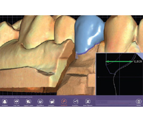
Актуальність. В ортопедичній стоматології на сучасному етапі її розвитку розроблено чимало технік препарування зубів під незнімні конструкції зубних протезів, однак не врахований стан пульпи у відповідь на препарування, внаслідок чого необґрунтованою є лікарська тактика щодо збереження чи екстирпації пульпи опорних зубів залежно від клінічного випадку. З огляду на загальноприйняті правила препарування зубів під незнімні ортопедичні конструкції існує значний відсоток ускладнень. Контроль глибини одонтопрепарування дає можливість провести цю маніпуляцію максимально раціонально та забезпечує ефективність протезування, виходячи зі стану вітальності препарованих зубів. Метa дослідження: визначення та контроль глибини препарування досліджуваних зубів із застосуванням техніки цифрового об’ємного сканування й алгоритмів цифрового графічного 3D редактора в системі CAD/CAM. Матеріали та методи. У статті наведено результати використання цифрового об’ємного сканування системи CAD/CAM та інтегрований алгоритм цифрового 3D моделювання для визначення та контролю глибини препарування зубів під незнімні ортопедичні конструкції. Результати. За результатами цифрового об’ємного сканування визначено робочу товщину одонтопрепарування в ділянці шийки зуба із формуванням різних видів шамфера в різних групах дослідження. Для визначення глибини препарування твердих тканин зуба було змодельовано штучну коронку в системі CAD — на цифровому об’ємному сканері NeWay (Open Technologies) та отримано дані про її товщину в ділянці уступу. Висновки. Метод цифрового об’ємного сканування для визначення та контролю глибини препарування твердих тканин зубів дає можливість запобігтити розвитку необоротних морфофункціональних змін в тканинах зубів, спричинених одонтопрепаруванням.

Background. In orthopedic dentistry at the present stage of its development, a chemical technique for preparing teeth for fixed structures of dentures has been developed; however, the state of the pulp in response to preparation is not taken into account. As a result, medical tactics for preserving or extirpating the pulp of abutment teeth, depending on the clinical case, are unreasonable. Given the generally accepted rules for preparing teeth for fixed orthopedic structures, there is a significant percentage of complications. Controlling the depth of tooth preparation makes it possible to carry out this manipulation as rationally as possible, and as a result to ensure the effectiveness of procedure, based on the state of vitality of the prepared teeth. The aim of the study is to determine and control the depth of the studied teeth preparation using digital volumetric scanning technology and algorithms of a digital graphic 3D editor in the CAD/CAM system. Materials and methods. The article presents the results of using digital volumetric scanning of the CAD/CAM system and an integrated algorithm for digital 3D modeling to determine and control the depth of teeth preparation for fixed orthopedic structures. Results. Based on the results of digital volumetric scanning, the working thickness of the tooth preparation in the area of the tooth neck was determined with the formation of various types of chamfer in different study groups. In order to determine the depth of the preparation of hard dental tissues, an artificial crown was modeled in the CAD system — digital volumetric scanner NeWay (Open Technologies) and data on its thickness in the area of the ledge were obtained. Conclusions. Method of digital volumetric scanning to determine and control the depth of preparation of hard dental tissues makes it possible to prevent the development of irreversible morpho-functional changes in the tissues of teeth caused by odontoreparation.


Ключевые слова

незнімне протезування зубів; CAD/CAM; шамфер; пришийковий уступ; штучна коронка

fixed dental prosthetics; CAD/CAM; chamfer; cervical ledge; artificial crown


Для ознакомления с полным содержанием статьи необходимо оформить подписку на журнал.


Список литературы

1. Gasiuk P.A., Radchuk V.B., Brekhlichuk P.P., Kalashnikov D.V., Zubchenko S.G. The influence of orthodontics preparation for the unfixed orthodontic constructions on the tooth pulp from the point of view of morphology. Intermedical Journal. 2015. 3(5). 39-43.
2. Dobrovolska O.V., Hasiuk N.V., Klytynska O.V., Zaliz-nyak M.S., Antonyshyn I.V. et al. State characteristics of the problem of oral cavity environmental system. Wiad. Lek. 2020. 73(5). 1037-1040.
3. Ram H.K., Shah R.J., Agrawal H.S. Evaluation of three different tooth preparation techniques for metal ceramic crowns by comparing preparation depths: an in vitro study. J. Indian Prosthodont. Soc. 2015. 15(2). 162-167. doi: 10.4103/0972-4052.159961.
4. Radchuk V.B., Hasiuk P.A., Hasiuk N.V. Morphofunctional changes of dental tissue innovative odontopreparation for metal-ceramic constructions. World of Medicine and Biology. 2017. 2(60). 100-103.
5. Pejčić A.S., Obradović R.R., Mirkovic D.S. The width of the attached gingiva and its variability in people with healthy periodontal status. Acta Stom. Naissi. 2017. 33(75). 1703-1717. doi: 10.5937/asn1775703P.
6. Radchuk V.B., Hasiuk N.V., Bozhyk S.S., Dzetsiukh T.I., Antonyshyn I.V. Initiating factors of complications development du-ring prosthetics of teeth with fixed prostheses. Wiad. Lek. 2021. 74(5). 1164-1168.
7. Roperto R.C., Oliveira M.P., Porto T.S., Ferreira L.A., Melo L.S., Akkus A. Can tooth preparation design affect the fit of CAD/CAM restorations? Compend. Contin. Educ. Dent. 2017. (3). e13-e17.
8. Šljivić K.Z., Nikolić M.D., Krunić N.S. Examining dentists’ attitudes about ethical principles in everyday practice. Acta Stom. Naissi. 2020. 36(82). 2067-2078. doi: 10.5937/asn2082067Z.
9. Shrestha B., Dunn L. The Declaration of Helsinki on Medical Research Involving Human Subjects: A Review of Seventh Revision. J. Nepal Health Res. Counc. 2020. 17(4). 548-552. doi: 10.33314/jnhrc.v17i4.1042.
10. Shillingburg H. Fundamentals of fixed prosthodontics. Third ed. Quintessence Publishing Co.; 2008. 557 p.
11. Hasiuk P., Vorobets A., Hasiuk N., Rosolovska S., Bodnarchuk I., Radchuk V. Sex differences of odontometrical indexes crowns of molars. Interventional Medicine & Applied Science. 2017. 9(3). 160-163. doi: 10.1556/1646.9.2017.08.
12. Zhang Y., Kelly J.R. Dental ceramics for restoration and metal veneering. Dent. Clin. North Am. 2017. 61(4). 797-819. doi: 10.1016/j.cden.2017.06.005.
13. Yang Y., Yang Z., Zhou J., Chen L., Tan J. Effect of tooth preparation design on marginal adaptation of composite resin CAD-CAM onlays. J. Prosthet. Dent. 2020. 124(1). 88-93. doi: 10.1016/j.prosdent.2019.08.010.
14. Ammoun R., Suprono M.S., Goodacre C.J., Oyoyo U., Carrico C.K., Kattadiyil M.T. Influence of Tooth Preparation Design and Scan Angulations on the Accuracy of Two Intraoral Digital Scanners: An in Vitro Study Based on 3-Dimensional Comparisons. J. Prosthodont. 2020. 29(3). 201-206. doi: 10.1111/jopr.13148.
15. Yu H.Y. Guided micro tooth preparation: from new strategies to new clinical practices. Zhonghua Kou Qiang Yi Xue Za Zhi. 2020. 55(10). 710-715. doi: 10.3760/cma.j.cn112144-20200627-00373.
16. Azarbal A., Azarbal M., Engelmeier R.L., Kunkel T.C. Marginal fit comparison of CAD/CAM crowns milled from two diffe-rent materials. J. Prosthodont. 2018. 27(5). 421-428. doi: 10.1111/jopr.12683.
17. Liang S., Yuan F. High-accuracy digital model design for full crown tooth preparation. Int. J. Comput. Dent. 2019. 22(4). 331-342.
18. Son K., Lee W.S., Lee K.B. Prediction of the learning curves of 2 dental CAD software programs. J. Prosthet. Dent. 2019. 121(1). 95-100. doi: 10.1016/j.prosdent.2018.01.004.
19. Hasiuk P., Hasiuk N., Kindiy D., Ivanchyshyn V., Kalashnikov D., Zubchenko S. Characteristics of cellular composition of periodontal pockets. Interv. Med. Appl. Sci. 2016. 8(4). 172-177. doi: 10.1556/1646.8.2016.4.5.
20. Liu C., Guo J., Gao J., Yu H. Computer-assisted tooth preparation template and predesigned restoration: a digital workflow. Int. J. Comput. Dent. 2020. 23(4). 351-362.

Вернуться к номеру