
Журнал "Гастроэнтерология" Том 57, №4, 2023
Вернуться к номеру
Ендоскопічна ультрасонографія в діагностиці захворювань органів травлення. Огляд клінічних випадків
Авторы: Степанов Ю.М., Пролом Н.В., Тарабаров С.О., Тітова М.В., Адамська І.М., Зеленюк О.В.
ДУ «Інститут гастроентерології НАМН України», м. Дніпро, Україна
Рубрики: Гастроэнтерология
Разделы: Клинические исследования
Версия для печати
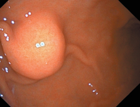
Ендоскопічне ультразвукове дослідження (ендоскопічна ультрасонографія, ЕУС) — це високотехнологічне ультразвукове дослідження, що одночасно поєднує в собі можливості ендоскопічної та ультразвукової діагностики захворювань шлунково-кишкового тракту, підшлункової залози, жовчних проток та печінки. Переваги ендоскопічного ультразвуку перед традиційним ультразвуковим дослідженням трансабдомінальним доступом полягають у тому, що ультразвуковий датчик через просвіт шлунково-кишкового тракту під візуальним контролем можна провести безпосередньо до досліджуваного об’єкта. ЕУС є методом вибору для дослідження підслизових утворень у верхніх відділах шлунково-кишкового тракту. Це найточніший метод для виявлення та діагностики підслизових утворень завдяки її високій чутливості, специфічності; використовується як наступний метод дослідження після ендоскопії та може надати інформацію про походження, розмір, межі, гомогенність утворень, а також щодо вибору методу лікування: ендоскопічне або хірургічне. Важливим застосуванням ЕУС є визначення стадії злоякісних новоутворень шлунково-кишкового тракту, оскільки це визначає лікування та прогнозує захворювання. У цьому допомагає компресійна еластографія в режимі реального часу, яка дозволяє аналізувати жорсткість тканин. Висока точність цього дослідження допомагає у диференційній діагностиці доброякісних та злоякісних новоутворень. У статті наведено випадки обстеження пацієнтів з підслизовими новоутвореннями шлунка в умовах ДУ «Інститут гастроентерології НАМН України».
Endoscopic ultrasound (EUS) is a high-tech ultrasound examination that simultaneously combines the options of endoscopic and ultrasound diagnosis of diseases of the gastrointestinal tract, pancreas, bile ducts and liver. The advantages of endoscopic ultrasound over traditional transabdominal ultrasound are that the ultrasound transducer can be guided directly through the lumen of the gastrointestinal tract to the object under visual control. The EUS is the method of choice for the study of submucosal lesions in the upper gastrointestinal tract. It is the most accurate method for the detection and diagnosis of submucosal formations due to its high sensitivity, specificity, is used as the next research method after endoscopy and can provide information about the origin, size, borders, homogeneity, as well as the choice of treatment method: endoscopic or surgical one. A more important application of EUS is the determination of the stage of malignant neoplasms of the gastrointestinal tract, as it determines the treatment and predicts the disease. And the real-time strain elastography, which allows analyzing the stiffness of tissues, helps in this. The high accuracy of this study allows for the differential diagnosis of benign and malignant neoplasms. The article presents cases of examination of patients with submucosal neoplasms of the stomach who were treated in the SI “Institute of Gastroenterology of the National Academy of Medical Sciences of Ukraine”.
ендоскопічне ультразвукове дослідження; еластографія; захворювання шлунково-кишкового тракту; підслизові новоутворення шлунка та дванадцятипалої кишки
endoscopic ultrasound; elastography; gastrointestinal diseases; submucosal neoplasms of the stomach and duodenum
Для ознакомления с полным содержанием статьи необходимо оформить подписку на журнал.
- Степанов Ю.М., Пролом Н.В., Коненко І.С., Тарабаров С.О., Недзвецька Н.В. Ендоскопічна ультразвукова сонографія в діагностиці патології шлунково-кишкового тракту. Гастроентерологія. 2021. Т. 55. № 3. С. 62-68. doi: https://doi.org/10.22141/2308-2097.55.3.2021.241590.
- Hocke M., Braden B., Jenssen C., Dietrich C.F. Present status and perspectives of endosonography 2017 in gastroenterology. Korean J Intern Med. 2018. № 33(1). P. 36-63. doi: 10.3904/kjim.2017.212.
- Iglesias-Garcia J., de la Iglesia-Garcia D., Lariño-Noia J., Dominguez-Muñoz J.E. Endoscopic Ultrasound (EUS) Guided Elastography. Diagnostics. 2023. № 13(10). P. 1686. https://doi.org/10.3390/diagnostics13101686.
- Endoscopic ultrasonography: An inside view / Simons-Lina–res C.R., Wander P., Vargo J., Chahal P. Cleve Clin J Med. 2020. № 87(3). P. 175-183. doi: 10.3949/ccjm.87a.19003.
- Competence in endosonographic techniques / P. Candoli, et al. Panminerva Med. 2019. № 61(3). P. 249-279. doi: 10.23736/S0031-0808.18.03570-X.
- Impact of endoscopic ultrasonography on diagnosis of pancrea–tic cancer / M. Kitano, et al. Gastroenterol. 2019. № 54(1). Р. 19-32. doi: 10.1007/s00535-018-1519-2.
- Endoscopic management of subepithelial lesions including neuroendocrine neoplasms: European Society of Gastrointestinal Endoscopy (ESGE) Guideline / P.H. Deprez, et al. Endoscopy. 2022. № 54(4). Р. 412-429. doi: 10.1055/a-1751-5742.
- Landi B., Palazzo L. The role of endosonography in submucosal tumours. Best Pract Res Clin Gastroenterol. 2009. № 23(5). Р. 679-701. doi: 10.1016/j.bpg.2009.05.009.
- Performance measures for ERCP and endoscopic ultrasound: a European Society of Gastrointestinal Endoscopy (ESGE) Quality Improvement Initiative / D. Domagk, et al. Endoscopy. 2018. № 50(11). Р. 1116-1127. doi: 10.1055/a-0749-8767.
- EUS-Guided Diagnosis of Gastric Subepithelial Lesions, What Is New? / T. Vasilakis, et al. Diagnostics (Basel). 2023. № 13(13). Р. 2176. doi: 10.3390/diagnostics13132176. PMID: 37443568.
- Endoscopic ultrasonography diagnosis of subepithelial lesions / M. Kida, dig. Endosc. Off. J Jpn Gastroenterol Endosc Soc. 2017. № 29. Р. 431-443.
- Diagnostic procedures for submucosal tumors in the gastrointestinal tract / L.G. Ponsaing, et al. World J Gastroenterol. 2007. Vol. 13(24). P. 3301-3310. doi: 10.3748/wjg.v13.i24.3301.
- Dhar J., Samanta J. The expanding role of endoscopic ultrasound elastography. Clin J Gastroenterol. 2022. № 15(5). Р. 841-858. doi: 10.1007/s12328-022-01662-0. PMID: 35789474.
- Maximizing the endosonography: The role of contrast harmo–nics, elastography and confocal endomicroscopy / Seicean A., Mostea–nu O., Seicean R. World J Gastroenterol. 2017. № 23(1) Р. 25-41. doi: 10.3748/wjg.v23.i1.25.
- Endoscopic Ultrasound-Guided Fine Needle Aspiration and Biopsy in Gastrointestinal Subepithelial Tumors / Pih G.Y., Kim D.H. Clin Endosc. 2019. № 52(4). Р. 314-320. doi: 10.5946/ce.2019.100.
- Sekine M., Asano T., Mashima H. The Diagnosis of Small Gastrointestinal Subepithelial Lesions by Endoscopic Ultrasound-Gui–ded Fine Needle Aspiration and Biopsy. Diagnostics (Basel). 2022. № 12(4). Р. 810. doi: 10.3390/diagnostics12040810.
- Endoscopic ultrasound elastography: Current status and future perspectives / X.W. Cui, et al. World J Gastroenterol. 2015. 21(47). Р. 13212-13224. PMID: 26715804; doi: 10.3748/wjg.v21.i47.13212.
- Efficacy and safety of endoscopic resection for small submucosal tumors originating from the muscularis propria layer in the gastric fundus / B. Li, et al. Surg Endosc. 2019. № 33(8). Р. 2553-2561. doi: 10.1007/s00464-018-6549-6.
- Nishida T., Kawai N., Yamaguchi S., Nishida Y. Submucosal tumors: comprehensive guide for the diagnosis and therapy of gastrointestinal submucosal tumors. Dig Endosc. 2013. № 25(5). Р. 479-89. doi: 10.1111/den.12149.
- Clinical course of suspected small gastrointestinal stromal tumors in the stomach / L.S. Ye et al. World J Gastrointest Surg. 2020. № 12(4). Р. 171-177. doi: 10.4240/wjgs.v12.i4.171. PMID: 32426096.
- Oda I., Suzuki H., Nonaka S., Yoshinaga S. Complications of gastric endoscopic submucosal dissection. Dig Endosc. 2013. Vol. 1. P. 71-78. doi: 10.1111/j.1443-1661.2012.01376.x.
- Pan W., Shi D. Band-assisted endoscopic mucosal resection for small (≤ 1.5 cm) submucosal tumors originating from the muscularis propria in the gastric fundus: a prospective study. Surg Endosc. 2023. № 37(3). Р. 1806-1812. doi: 10.1007/s00464-022-09688-8.
- Franco M.C., Schulz R.T., Maluf-Filho F. Opinion: How to mana–ge subepithelial lesions of the upper gastrointestinal tract? World J Gastrointest Endosc. 2015. № 7(18). Р. 1262-7. doi: 10.4253/wjge.v7.i18.1262.
- Hu J., Ge N., Wang S., Guo J., Liu X., Wang G., Sun S. Direct endoscopic full-thickness resection for submucosal tumors with an intraluminal growth pattern originating from the muscularis propria layer in the gastric fundus. BMC Gastroenterol. 2020. № 20(1). Р. 70. doi: 10.1186/s12876-020-01215-0.