Журнал «Боль. Суставы. Позвоночник» Том 14, №4, 2024
Вернуться к номеру
Результати експериментального моделювання напружень на фіксатори при металоостеосинтезі черезвертлюгових переломів
Авторы: Калашніков А.В., Сабарна Ю.Х.М.
ДУ «Інститут травматології та ортопедії НАМН України», м. Київ, Україна
Рубрики: Ревматология, Травматология и ортопедия
Разделы: Клинические исследования
Версия для печати
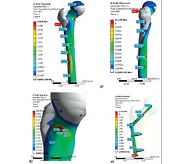
Актуальність. У розвинутих країнах світу при лікуванні переломів проксимального відділу стегнової кістки широко впроваджують малоінвазивні технології застосування проксимального стегнового стрижня. Проте нами не знайдено літературних даних щодо напружень на блокований інтрамедулярний стрижень залежно від типу перелому за класифікацією Aсоціації остеосинтезу та варіантів його дистального блокування. Мета дослідження: провести аналіз напружень на різні металеві фіксатори при виконанні остеосинтезу з приводу черезвертлюгових переломів типу А1. Матеріали та методи. Використовували макет стегнової кістки, у який імплантовано фіксуючі елементи. Для фіксації відламків застосовували 2 варіанти фіксаторів — динамічна кульшова пластина (DHS, 1-й варіант) і проксимальний стегновий стрижень (PFN, 2-й варіант), які забезпечують оптимальні біомеханічні й біологічні умови для зрощення переломів. Розрахунки напружено-деформованого стану методом кінцевих елементів проводили для інтактної моделі з обома варіантами фіксаторів, а потім з фіксаторами при черезвертлюгових переломах типу А1 і варіантами дистального блокування (без блокування, 1 гвинтом, 2 гвинтами). Мінімальне напруження на металеві фіксатори в їх проксимальних відділах визначали при застосуванні DHS-пластини та PFN-стрижня у варіанті без застосування гвинтів для дистального блокування. Результати. Отримані результати вірогідно (p ≤ 0,05) відрізнялися від даних при застосуванні PFN-стрижня з дистальним блокуванням 1 чи 2 гвинтами. Напруження на дистальний відділ металевих фіксаторів не виникало при застосуванні PFN-стрижня у варіанті без застосування гвинтів для дистального блокування. На відміну від цього при застосуванні DHS-пластини напруження збільшувалося до максимальних показників і становило 57,18 МПа. Визначено, що максимальна, але адекватна мікрорухливість спостерігалася при застосуванні моделі PFN-стрижня у варіанті без застосування гвинтів для дистального блокування. Ці мікрорухи сприятимуть поліпшенню репаративного остеогенезу черезвертлюгових переломів типу А1. Занадто міцна фіксація при застосуванні DHS-пластини (1,95 мм) може призвести до порушення репаративного остеогенезу. Висновки. Проведене дослідження визначає диференційований підхід до лікування хворих із черезвертлюговими переломами стегнової кістки, що покращить ефективність надання медичної допомоги цій тяжкій категорії хворих.
Background. In the world’s developed countries, minimally invasive technologies for using the proximal femoral rod are widely implemented in treating fractures of the proximal femur. However, we did not find literature data on the stresses on the blocked intramedullary rod depending on the type of fracture due to АО (Association of Osteosynthesis) and variants of its distal blocking. The study aimed to carry out biomechanical modeling of stresses on various metal fixators in osteosynthesis for transtrochanteric fractures of type A1. Materials and methods. We used a model of the femur, in which fixing elements were implanted. Two types of fixators were used to fix the fragments — the DHS (dynamic hip screw) plate (option 1) and the proximal femoral nail (PFN, option 2) rod, which provide optimal biomechanical and biological conditions for fracture union. Calculations of stress-strain state by the finite element method were performed for the intact model with both types of fixators and then with fixators for transtrochanteric fractures type A1 and options for distal locking (without locking, 1 screw, 2 screws). It was determined that the minimum stress on the metal fasteners in their proximal parts was determined when using the DHS plate and the PFN rod in the version without the use of screws for distal locking. Results. Our results were significantly (p ≤ 0.05) different from the data when using a PFN rod with distal locking with 1 or 2 screws. There was no stress on the distal part of the metal retainers when the PFN rod was applied in the variant without the use of screws for distal locking. In contrast, the stress when the DHS plate was applied increased to maximum values of 57.18 MPa. It was determined that maximum but adequate micromobility was observed when using the PFN rod model in the version without the use of screws for distal locking. These micromovements will contribute to the improvement of reparative osteogenesis of patients with type A1 transtrochanteric fractures. Fixation that is too strong when using a DHS plate (1.95 mm) can lead to impaired reparative osteogenesis. Conclusions. The research will make it possible to determine a differentiated approach to treating patients with transtrochanteric fractures of the femur. It will improve the effectiveness of providing medical care to this severe category of patients.
комп’ютерне моделювання; переломи проксимального відділу стегнової кістки; напруження; металоостеосинтез
computer modeling; proximal femur fractures; stress; metallo-osteosynthesis
Для ознакомления с полным содержанием статьи необходимо оформить подписку на журнал.
- Поворознюк В.В., Григор’єва Н.В., Корж М.О. та ін. Епідеміологія переломів проксимального відділу стегнової кістки в Україні: результати двох ретроспективних досліджень. Ортопедия, травматология и протезирование. 2016;(4):68-74. doi: 10.15674/0030-59872016468-74.
- Saltvedt I, Prestmo A, Einarsen E, Johnsen LG, Helbostad JL, Sletvold O. Development and delivery of patient treatment in the Trondheim Hip Fracture Trial. A new geriatric in-hospital pathway for elderly patients with hip fracture. BMC Res. Notes. 2012;(5):355. doi: 10.1186/1756-0500-5-355.
- Kanis JA, Odén A, McCloskey EV, Johansson H, Wahl DA, Cooper C. IOF Working Group on Epidemiology and Quality of Life. A systematic review of hip fracture incidence and probability of fracture worldwide. Osteoporosis international. 2012;23(9):2239-2256. doi: 10.1007/s00198- 012-1964-3.
- Choy WS, Kim KJ, Lee SK, Yang DS, Jeung SW, Choi HG, Park HJ. Surgical treatment of pathological fractures occurring at the proximal femur. Yonsei Medical Journal. 2015;56(2):460-465. doi: 10.3349/ymj.2015.56.2.460.
- Walter N, Szymski D, Kurtz S, Alt V, Lowenberg DW, Lau E, Rupp M. Factors associated with morta–lity after proximal femoral fracture. Journal of Orthopaedics and Traumatology. 2023;(24):31. doi: 10.1186/s10195-023-00715-5.
- Піонтковський В.К., Денисюк Б.С., Малевич Ю.М., Каштан Ю.М., Циплінський Ю.І., Златів В.П., Столярський Н.І. Наш досвід малоінвазивного остеосинтезу при черезвертлюжних переломах стегнової кістки. Травма. 2012;13(4):140-142.
- Smektala R, Endres HG, Dasch B, Maier C, Trampisch HJ, Bonnaire F, Pientka L. The effect of time-to-surgery on outcome in elderly patients with proximal femoral fractures. BMC musculoskeletal disorders. 2008;(9):171. doi: 10.1186/1471-2474-9-171.
- Kijima H, мYamada S, Konishi N, Kubota H, Tazawa H, Tani T et al. The Reliability of Classifications of Proximal Femoral Fractures with 3-Dimensional Computed Tomography: The New Concept of Comprehensive Classification. Advances in Orthopedics. 2014:1. doi: 10.1155/2014/359689.
- Fischer H, Maleitzke T, Eder C, Ahmad S, Stockle U, Braun KF. Management of proximal femur fractures in the elderly: current concepts and treatment options. Eur J Med Res. 2021;26:86. doi: 10.1186/s40001-021-00556-0.
- Ding K, Zhu Y, Zhang Y, Li Y, Wang H, Li J, Chen W, Zhang Q. Proximal femoral bionic nail — a novel internal fixation system for the treatment of femoral neck fractures: a finite element analysis Front. Bioeng Biotechnol. 2023:11:1297507. doi: 10.3389/fbioe.2023.1297507.
- Sadic S, Custovic S, Jasarevic M, Fazlic M, Smajic N, Hrustic A, Vujadinovic A, Krupic F. Proximal femoral nail antirotation in treatment of fractures of proximal femur. Medicinski arhiv. 2014;68(3):173-177. doi: 10.5455/medarh.2014.68.173-177.
- Liu H, Yuan B, Yu T, Ren G, Zhang Y, Wu Y, Liu Y, Peng C, Wu D Accurate fixation of complicated comminu–ted femur fracture with customized LCP refe–rencing a life-size 3D-printed model: Annals of Translational Medicine 2020;8(7):505. doi: 10.21037/atm.2020.03.115.
- Маланчук В.О., Крищук М.Г., Копчак-Маланчук А.В. Імітаційне комп’ютерне моделювання в щелепно-лицевій хірургії. Київ: Асканія, 2013. С. 45-49.
- Никифоров Р.Р., Куценко С.Н., Костандов Ю.А. Механико-математическая модель системы металлоостеосинтеза и расчет ее напряженно-деформированного состояния. Травма. 2013;14(3):43-51.
- Mostafa IA, El-Sayed Abdel-Aal SA, Yassin MO. Proximal Femoral Plate Versus Proximal Femoral Nailing Fixation for Treatment of Unstable Intertrochanteric Fe–moral Fractures. Al-Azhar International Medical Journal. 2024;5(193): 206. doi: 10.58675/2682-339X.2282.
- Katnic I, Orlandic MI. Fundamentals of Biomedical Statistics. Stud Health Technol Inform. 2020;274:111-121. doi: 10.3233/SHTI200671.
- Molinari G, Emiliani N, Cercenelli L, Bortolani B, Gironi C, Javier I, Presutti Fl, Marcelli E. Assessment of a novel patient-specific 3D printed multi-material simulator for endoscopic sinus surgery. Front Bioeng Biotechnol. 2022;17(10):974021. doi: 10.3389/fbioe.2022.974021.
- Zaczyk M, Jasińska-Choromańska D. Contact phenomena modeling in biological structures on the example of the implant-bone. Latin American Journal of Solids and Structures. 2019:16 (1 Thematic Section): e172. doi: 10.1590/1679-78254955.
- Tretiakow D, Tesch K, Markiet K. Numerical ana–lysis of the ostiomeatal complex aeration using the CFD method. Scientific Reports. 2023;13:1-11. doi: 10.1038/s41598-023-31166-x.
- Galbusera F, Cina A, Panico M, Albano D, Messina C. Image-based biomechanical models of the musculoskeletal system. European Radiology Experimental. 2020;4:49. doi: 10.1186/s41747-020-00172-3.

