
Журнал «Травма» Том 26, №4, 2025
Вернуться к номеру
Ефективність і ризики зовнішньої фіксації при переломах великогомілкової кістки: метааналіз методів Ілізарова і Тейлора
Авторы: Суворов В.Л. (1), Кулик Ю.А. (1), Дем’ян Ю.Ю. (1), Полулях Д.М. (1), Євлантьєва Т.А. (1), Ларкевич О.Г. (1), Козік Є.В. (1), Карпінська О.Д. (2)
(1) - ДУ «Інститут травматології та ортопедії НАМН України», м. Київ, Україна
(2) - ДУ «Інститут патології хребта та суглобів імені професора М.І. Ситенка НАМН України», м. Харків, Україна
Рубрики: Травматология и ортопедия
Разделы: Клинические исследования
Версия для печати
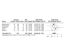
Актуальність. Переломи великогомілкової кістки залишаються однією з найпоширеніших і водночас найскладніших травм опорно-рухового апарату. Їх лікування часто ускладнюється значним пошкодженням м’яких тканин, відкритими ранами, нестабільністю відламків та ризиком інфекційних ускладнень. У таких випадках методи зовнішньої фіксації є важливим інструментом стабілізації, що дозволяє забезпечити контроль над положенням відламків при мінімальній інвазивності. Серед основних методів зовнішньої фіксації при лікуванні переломів великогомілкової кістки найбільш широке застосування отримали класичний апарат Ілізарова та сучасні гексаподні системи, як-от просторова рамка Тейлора (Taylor Spatial Frame, TSF). Мета: провести систематичне порівняння ефективності та безпечності методу Ілізарова та просторової рамки Тейлора у лікуванні переломів великогомілкової кістки з урахуванням часу консолідації перелому, частоти ускладнень, часу зрощення та ступеня корекції деформацій. Матеріали та методи. Пошук джерел проводився в базах даних PubMed, Semantic Scholar та Scopus за період з 2014 до 2025 року. За всіма базами було виявлено 105 робіт. Усі включені дослідження мали ретроспективний когортний дизайн. Рандомізованих контрольованих досліджень, що відповідали критеріям PICO, виявлено не було. Результати. Загальна кількість спостережень становила 723 пацієнти (338 осіб — метод Ілізарова, 385 — Тейлора). Не визначено доказів суттєвої різниці в кількості відкритих і закритих переломів. Незважаючи на високу гетерогенність (I2 = 92 %) досліджень, не визначено значущої різниці між системами щодо термінів консолідації переломів. Терміни загоєння сильно варіювалися залежно від місця перелому, його складності, первинного стану. Група Ілізарова мала трохи вищий, але не значущий індекс фіксації. Різниця в ризику інфекцій між методами не є статистично значущою. Про величину залишкової деформації повідомлено у двох дослідженнях. Кут залишкової деформації великогомілкової кістки в групі Ілізарова був на 0,7° більшим, ніж у групі TSF, різниця статистично значуща. Висновки. За даними проведеного метааналізу, при порівнянні ефективності та безпечності методу Ілізарова та просторової рамки Тейлора у лікуванні переломів великогомілкової кістки не було виявлено статистично значущої різниці між цими системами ані за термінами зрощення перелому, ані за кількістю ускладнень. Водночас рамка Тейлора показала вірогідно кращі результати щодо остаточної деформації кінцівки. Слід відзначити, що накопичений клінічний досвід і вдосконалення рекомендацій щодо застосування обох методів фіксації залежно від типу, локалізації та складності перелому майже знівелювали відмінності в частоті ускладнень. Варто зауважити, що хоча деякі автори наводили дані про типи та складність переломів великогомілкової кістки, узагальнюючі результати публікувались без конкретної прив’язки до цих характеристик. Тому неможливо зробити остаточні висновки про перевагу тієї чи іншої системи в окремих клінічних випадках. З огляду на значну індивідуальну варіативність переломів питання ефективності та безпечності методів лікування можуть бути остаточно вирішені лише за умов наявності достатньо великого обсягу клінічних даних.
Background. Fractures of the tibia remain one of the most common and at the same time most complex injuries of the musculoskeletal system. Their treatment is often complicated by significant soft tissue damage, open wounds, instability of fragments, and the risk of infectious complications. In such cases, external fixation methods are an important stabilizing tool that allows control over the position of fragments with minimal invasiveness. Among the main methods for external fixation in the treatment of tibial fractures, the classic Ilizarov apparatus and modern hexapod systems, such as the Taylor spatial frame (TSF), are most widely used. The purpose was to conduct a systematic comparison of the efficacy and safety of the Ilizarov method and the TSF in the treatment of tibial fractures, taking into account the time of fracture consolidation, the frequency of complications, the time of union, and the degree of deformity correction. Materials and methods. The search was conducted in the PubMed, Semantic Scholar, and Scopus databases from 2014 to 2025. A total of 105 studies were identified in all databases. All included studies had a retrospective cohort design. No randomized controlled trials meeting the PICO criteria were found. Results. The total number of observations was 723 patients (338 individuals using the Ilizarov method, 385 using the Taylor method). There was no evidence of a significant difference in the number of open and closed fractures. Despite the high heterogeneity (I2 = 92 %) of the studies, no significant difference was found between the systems in terms of fracture consolidation times. Healing times varied greatly depending on the location of the fracture, its complexity, and the initial condition. The Ilizarov group had a slightly higher, but not significant, fixation index. The difference in the risk of infection between the methods is not statistically significant. The magnitude of residual deformity was reported in two studies. The angle of residual deformity of the tibia in the Ilizarov group was 0.7° greater than in the TSF group, the difference is statistically significant. Conclusions. According to the meta-analysis, when comparing the efficacy and safety of the Ilizarov method and the TSF in the treatment of tibial fractures, no statistically significant differences were found between these systems in terms of fracture healing time or the number of complications. At the same time, the Taylor frame showed significantly better results in terms of final limb deformity. It should be noted that the accumulated clinical experience and improvement of recommendations for the use of both fixation methods, depending on the type, location, and complexity of the fracture, have almost eliminated the differences in the frequency of complications. It should be noted that although some authors provided data on the types and complexity of tibial fractures, the summary results were published without specific reference to these characteristics. Therefore, it is impossible to draw definitive conclusions about the superiority of one system or another in individual clinical cases. Given the significant variability of fractures, the effectiveness and safety of treatment methods can only be reliably determined if a sufficiently large amount of clinical data is available.
перелом великогомілкової кістки; просторова рамка Тейлора; система Ілізарова; метааналіз
tibial fracture; Taylor spatial frame; Ilizarov system; meta-analysis