Інформація призначена тільки для фахівців сфери охорони здоров'я, осіб,
які мають вищу або середню спеціальну медичну освіту.

Підтвердіть, що Ви є фахівцем у сфері охорони здоров'я.


Всесвітній день боротьби із запальними захворюваннями кишечника
день перший
день другий

Коморбідний ендокринологічний пацієнт

Всесвітній день боротьби із запальними захворюваннями кишечника
день перший
день другий

Коморбідний ендокринологічний пацієнт

Международный эндокринологический журнал Том 16, №4, 2020

Вернуться к номеру

Correlation of vitamin D level with thyroid status and TSH antibody titers in patients with Graves’ disease

Авторы: V.I. Pankiv(1), T.Yu. Yuzvenko(1), S.M. Koval(2), K. Singh(1), I.V. Pankiv(3), Tarun Sehgal(4), O.M. Lytvinova(5)
(1) — Ukrainian Research and Practical Centre of Endocrine Surgery, Transplantation of Endocrine Organs and Tissues of the Ministry of Health of Ukraine, Kyiv, Ukraine
(2) — L.T. Mala National Institute of Therapy of the NAMN Ukraine, State Institution, Kharkiv, Ukraine
(3) — Bukovinian State Medical University, Chernivtsi, Ukraine (4) — Dr B.R. Ambedkar National Institute of Technology, Jalandhar, Punjab, India
(5) — National University of Pharmacy, Kharkiv, Ukraine

Рубрики: Эндокринология

Разделы: Клинические исследования

Версия для печати


Резюме

Актуальність. У багатьох дослідженнях встановлено зв’язок дефіциту вітаміну D з автоімунними захворюваннями. Доведено, що рівень вітаміну D у пацієнтів з автоімунними захворюваннями щитоподібної залози, включаючи хворобу Грейвса, був нижчим, ніж у пацієнтів із неавтоімунними тиреоїдними захворюваннями, наприклад токсичним вузловим зобом. Водночас в окремих статтях повідомляють про відсутність такої залежності між рівнем вітаміну D та автоімунними захворюваннями щитоподібної залози. Мета дослідження: порівняти рівень вітаміну D у сироватці крові в пацієнтів із хворобою Грейвса та осіб контрольної групи, оцінити співвідношення вмісту вітаміну D із функціональним станом щитоподібної залози і титром антитіл до рецептора тиреотропного гормону (ТТГ). Матеріали та методи. Під спостереженням перебували 48 пацієнтів із хворобою Грейвса та 24 здорові особи конт­рольної групи. Усім обстеженим проводили гормональні дослідження, які містили рівень ТТГ сироватки, тироксину, вільного трийодтироніну, паратиреоїдного гормону, а також визначали рівень іонізованого кальцію, 25-гідроксивітаміну D (25(OH)D), титру антитіл до рецептора ТТГ. Результати. У пацієнтів із хворобою Грейвса спостерігався вірогідно нижчий рівень 25(OH)D (16,3 ± 1,4 нг/мл) порівняно з показниками контрольної групи (22,8 ± 1,6 нг/мл) (p = 0,024). Рівні ТТГ і титри антитіл до рецептора ТТГ вірогідно відрізнялися між групою осіб із хворобою Грейвса з дефіцитом вітаміну D (25(OH)D < 20 нг/мл) та групою хворих із хворобою Грейвса без дефіциту вітаміну D (25(OH)D ≥ 20 нг/мл). Обсяг щитоподібної залози істотно не відрізнявся між цими групами хворих. Рівень вітаміну D у сироватці крові вірогідно взаємопов’язаний із вмістом ТТГ і титром антитіл до рецептора ТТГ у пацієнтів із хворобою Грейвса. Висновки. Рівень вітаміну D у сироватці крові вірогідно нижчий у пацієнтів із хворобою Грейвса. У цих пацієнтів встановлено вірогідну кореляцію між вмістом вітаміну D і рівнями ТТГ й антитіл до рецептора ТТГ.

Актуальность. Во многих исследованиях установлена связь дефицита витамина D с аутоиммунными заболеваниями. Доказано, что уровень витамина D у пациентов с ауто­иммунными заболеваниями щитовидной железы, включая болезнь Грейвса, был ниже, чем у пациентов с неаутоиммунными тиреоидными заболеваниями, например токсическим узловым зобом. В то же время в отдельных статьях сообщают об отсутствии такой зависимости между уровнем витамина D и аутоиммунными заболеваниями щитовидной железы. Цель исследования: сравнить уровень витамина D в сыворотке крови у пациентов с болезнью Грейвса и лиц контрольной группы, оценить соотношение содержания витамина D с функциональным состоянием щитовидной железы и титром антител к рецептору тиреотропного гормона (ТТГ). Материалы и методы. Под наблюдением находились 48 пациентов с болезнью Грейвса и 24 здоровых лица контрольной группы. Всем обследованным проводили гормональные исследования, содержащие уровень ТТГ сыворотки, свободного тироксина, свободного трийодтиронина, паратиреоидного гормона, а также определяли уровень ионизированного кальция, 25-гидроксивитамина D (25(OH)D), титр антител к рецептору ТТГ. Результаты. У пациентов с болезнью Грейвса наблюдался достоверно более низкий уровень 25(OH)D (16,3 ± 1,4 нг/мл) по сравнению с показателями контрольной группы (22,8 ± 1,6 нг/мл) (p = 0,024). Уровни ТТГ и титры антител к рецептору ТТГ достоверно отличались между группой лиц с болезнью Грейвса с дефицитом витамина D (25(OH)D < 20 нг/мл) и группой больных с болезнью Грейвса без дефицита витамина D (25(OH)D ≥ 20 нг/мл). Объем щитовидной железы существенно не отличался между этими группами больных. Уровень витамина D в сыворотке крови достоверно взаимосвязан с концентрацией ТТГ и титром антител к рецептору ТТГ у пациентов с болезнью Грейвса. Выводы. Уровень витамина D в сыворотке крови достоверно ниже у пациентов с болезнью Грейвса при сравнении с показателями контрольной группы. У этих пациентов установлена достоверная корреляция между содержанием витамина D и уровнями ТТГ и антител к рецептору ТТГ.

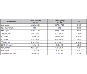
Background. Various studies have shown the association of vitamin D deficiency with autoimmune diseases. Studies have found that vitamin D levels in patients with autoimmune thyroid diseases including Graves’ disease were lower than that in patients with non-autoimmune thyroid diseases such as toxic nodular goiter. Some studies have reported no such relationship between vitamin D level and autoimmune thyroid di­seases. The purpose of the study: to compare serum vitamin D level in patients with Graves’ disease versus age and sex matched controls, to assess the correlation of vitamin D with thyroid status and thyrotropin receptor antibody titers. Materials and methods. 48 patients with Graves’ disease and 24 age and sex matched healthy individuals were recruited. Hormonal investigations that included serum thyroid stimulating hormone (TSH), free thyroxine (fT4), free triiodothyronine (fT3), as well as calcium, 25-hydroxyvitamin D (25(OH)D), parathyroid hormone (PTH), thyroid stimulating hormone receptor antibody (TSH-Ab) were done for all subjects. Results. The patients with Graves’ disease had significantly lower 25(OH)D levels (16.3 ± 1.4 ng/ml) as compared to control subjects (22.8 ± 1.6 ng/ml) (p = 0.024). TSH levels and TSH-Ab titers differed significantly between vitamin D deficient Graves’ disease group (25(OH)D < 20 ng/ml) and vitamin D non deficient Graves’ disease group (25(OH)D ≥ 20 ng/ml). Thyroid volume did not differ significantly between these groups. Serum vitamin D level correlated significantly with TSH and TSH-Ab titers in patients with Graves’ disease. Conclusions. Serum vitamin D levels are significantly lower in patients with Graves’ disease. Significant correlation between vitamin D and TSH and TSH-Ab titers was found in these patients.


Ключевые слова

хвороба Грейвса; вітамін D; антитіла до рецептора ТТГ

болезнь Грейвса; витамин D; антитела к рецептору ТТГ

Graves’ disease; vitamin D; TSH-Ab titers

Introduction

Graves’ disease is one of the common autoimmune thyroid disease encountered in routine clinical practice. In recent years, the extraskeletal effects of vitamin D have been extensively studied. Vitamin D deficiency is linked to a variety of autoimmune disorders, including autoimmune thyroid disease [1]. Several studies suggest that individuals with Graves’ disease have lower vitamin D levels than the general population [2–4]; however, data on the relationship between the levels of vitamin D and clinical parameters in Graves’ disease or therapeutic issues [5] are limited. A few studies have analyzed the association between serum vitamin D levels and Graves’ disease, and available data remain inconclusive. Various studies have shown the association of vitamin D deficiency with autoimmune diseases. In past, studies have found that vitamin D levels in patients with autoimmune thyroid diseases including Graves’ disease were lower than that in patients with non-autoimmune thyroid diseases [6, 7].
To understand the role of vitamin D in Graves’ disease, it is important to determine whether lower vitamin D levels contribute to the development of Graves’ disease. There are some animal data supporting this hypothesis. BALB/cJ mice given a vitamin D-deficient diet had lower preimmunization thyroxine levels and were more prone to developing persistent hyperthyroidism following immunization with the TSH receptor compared to mice fed regular chow. No differences in the TSH-Ab levels were observed, suggesting that vitamin D directly modulated thyroid function in this animal model [8]. The lack of a significant association between vitamin D and TSH receptor antibody would support the hypothesis that vitamin D deficiency might have a direct effect on the thyroid gland [9]. However, as all clinical studies that address this question have been cross-sectional in design, it is impossible to conclude whether the vitamin D status is directly involved in the pathogenesis or a consequence of the disease. 
Hence, the relationship between vitamin D and Graves’ disease is still unresolved. 
We purposed to compare serum vitamin D level in patients with Graves’ disease versus age and sex matched controls and to find associations of vitamin D status and Graves’ disease and ascertain any correlation of serum vitamin D level with various thyroid related parameters.

Materials and methods

A total of 48 patients (34 females and 14 males) with Graves’ disease were recruited to this cross-sectional study. Graves’ disease was diagnosed by clinical, hormonal evidence of hyperthyroidism (suppressed thyroid stimulating hormone (TSH) level with elevated free triiodothyronine (fT3), and free thyroxine (fT4) levels. Forty-four age and sex matched healthy individuals (21–55 years) meeting the exclusion criteria were taken as controls. To avoid influence of seasonal variation on vitamin D, we recruited controls within the same month (± 1 month) as that of cases. 
Exclusion criteria included patients with liver, renal, metabolic bone, parathyroid disorders, any autoimmune diseases, history of intake of calcium and vitamin D supplements within preceding 3 months, current or recent use of steroids, antitubercular drugs, antiepileptics, ketoconazole, heparin, oral contraceptives and diuretics, pregnant, and lactating women. All study participants underwent a detailed history enquiry and clinical examination. 
We had divided the Graves’ disease cohort broadly into two groups: vitamin D deficiency group (25(OH)D < 20 ng/ml) and vitamin D insufficiency group (< 30 ng/ml 25(OH)D ≥ 20 ng/ml). 
After an overnight fast, blood samples were collected for measurements of TSH, fT3, fT4, 25(OH)D, calcium and parathyroid hormone (PTH) and thyrotropin receptor antibody (TSH-Ab). Serum TSH, fT3, fT4, PTH, and 25(OH)D were measured using electrochemiluminescene immunoassay kits. 
The thyroid gland correct volume was obtained using ellipsoid formula after measuring maximum the longitudinal (L), anteroposterior (AP), and transverse (T) axis of both lobes. Both lobe volumes were added to get the total volume of the gland. Written informed consent was taken from all study participants. 
Descriptive statistical methods such as mean and standard deviation were applied to summarize continuous variables. Categorical data were summarized as percentages or proportions. Normality distribution of all parameters was checked using Shapiro-Wilk test. Categorical variables were compared using Chi-square test. Mann-Whitney U test and independent t tests were performed to compare means between two groups as required. Kruskal Wallis test was used for comparison of means between three or more groups. Pearson’s correlation coefficient was used to analyze correlation between different parameters. A p-value of less than 0.05 was considered statistically significant. 
This case-control study was performed with the Institutional Review Board protocol approval 25.02.2019 (number 7/2019) in Ukrainian Scientific and Practical Center for Endocrine Surgery, Transplantation of Endocrine Organs and Tissues of the Ministry of Health of Ukraine, between May — September 2019. 
The study was conducted as part of a comprehensive scientific research «Optimization of prevention, diagnosis and treatment of diabetes mellitus on the background of comorbid pathology, taking into account the effects of iodine and vitamin D deficiency» (Health Ministry of Ukraine registration 0120U000218).

Results

A total of 48 patients with Graves’ disease and 24 age and sex matched controls were included in the study. The baseline characteristics of the study population are summarized in table 1. The mean age of patients with Graves’ disease was 36.45 ± 8.60 years as compared to mean age of 33.75 ± 7.90 years among controls (p = 0.105). Graves’ disease patients had significantly suppressed TSH levels, elevated fT3 and elevated fT4 in comparison to controls (p < 0.001). The mean thyroid volume was significantly higher among Graves’ disease patients (29.2 ± 8.3 cm3) as compared to controls (12.5 ± 2.7 cm3) (p < 0.05).
It was seen that mean serum 25(OH) D levels were significantly lower among Graves’ disease patients (16.3 ± ± 1.4 ng/ml) versus controls (22.8 ± 1.6 ng/ml) (p = 0.024). The serum calcium and PTH levels did not vary significantly among Graves’ disease and control groups. 
The comparison of different parameters between groups with and without vitamin D deficiency are summarized in table 2. There wasn’t significant difference among these two subgroups of Graves’ disease patients with regard to mean serum TSH and serum TSH-Ab levels. 

Discussion

We found that serum mean 25(OH) D levels were significantly lower in Graves’ disease patients as compared to controls. 
One of the first studies that reported widespread vitamin D deficiency among Graves’ disease patients was reported by H. Yamashita et al. [10]. The mean level of vitamin D in men and women Graves’ disease subjects were 41.3 ± 15.0 and 31.8 ± 13.3 nmol/l, respectively in their cohort. In 2013, T. Yasuda et al. reported significantly lower mean 25(OH)D level among new onset Graves’ disease female patients versus controls and suggested toward association of vitamin D and Graves’ disease [3]. The findings of low vitamin D status in Graves’ disease of our study is in agreement to reports from previous studies [11–13]. In a recently published study from Sweden, authors have reaffirmed the finding of lower vitamin D levels in Graves’ disease [9]. 
However, some studies have not reported similar finding of low vitamin D status among Graves’ disease than control population [14]. In a meta-analysis to study the association of vitamin D and Graves’ disease, authors concluded that the patients with Graves’ disease were more likely to be deficient in vitamin D compared to the controls with a high heterogeneity [15]. 
In our study, we found statistical difference in terms of TSH and serum TSH antibody titers between groups with and without vitamin D deficiency. Only one previous study has looked into the difference of thyroid related parameters between two Graves’ disease groups stratified on basis of mean serum vitamin D level (25(OH)D < 20 ng/ml group versus 25(OH)D > 20 ng/ml group) [14]. The authors noted that the two groups had no significant difference in terms of thyroid hormone status. They also reported that 100% of their Graves’ disease patients were serum TSH antibody titers positive when 25(OH)D level < 20 ng/ml as compared to 37.5 % of patients were serum TSH antibody titers positive when 25(OH)D level > 20 ng/ml. 
Among our cohort, patients with lowest vitamin D levels (group with 25(OH)D < 20 ng/ml) had significantly the highest mean serum TSH antibody titers. Other authors [4] have noted lower mean 25(OH)D levels among serum TSH antibody titers positive Graves’ disease patients in comparison to serum TSH antibody titers negative Graves’ disease patients. Our results show that serum vitamin D level correlated with TSH level and serum TSH antibody titers in Graves’ disease. Yasuda T. et al. [3] showed no association between serum 25(OH)D levels and thyroid function tests or serum TSH antibody titers. Recently published study [14] also did not report any correlation of serum vitamin D level with thyroid hormones and thyroid auto-antibody titers.
However, in contrast to these findings, two studies have showed significant inverse correlation between vitamin D levels and serum TSH antibody titers in Graves’ disease [5, 11]. From review of above findings, it seems that thyroid hormones are affected by vitamin D status in Graves’ disease. Nevertheless, the relationship between vitamin D level and antibody titers need further clarification, though at present most of studies fail to report any significant correlation between these two parameters [14].
Our study has few limitations. We did not assess daily total dietary calcium, vitamin D, and phytate intake that may have affected vitamin D status. Serum 25(OH)D assay was done only once for each person. Being a cross-sectional study causality between vitamin D deficiency and risk of Graves’ disease cannot be made. Our sample size was small, and hence, future large scale studies are essential to further clarify the findings.

Conclusions

Vitamin D levels are significantly lower in patients with Graves’ disease. Correlation was observed between vitamin D and TSH level and serum TSH antibody titers in Graves’ disease. Vitamin D deficiency is significantly associated with Graves’ disease.
Conflicts of interests. Authors declare the absence of any conflicts of interests and their own financial interest that might be construed to influence the results or interpretation of their manuscript.

Список литературы

  1. Villa A., Corsello A., Cintoni M. et al. Effect of vitamin D supplementation on TSH levels in euthyroid subjects with autoimmune thyroiditis. Endocrine. 2020;10.1007/s12020-020-02274-9. doi: 10.1007/s12020-020-02274-9.
  2. Krysiak R., Kowalcze K., Okopień B. The effect of vitamin D on thyroid autoimmunity in euthyroid.men with autoimmune thyroiditis and testosterone deficiency. Pharmacol. Rep. 2019. 71(5). Р. 798-803. doi: 10.1016/j.pharep.2019.04.010.
  3. Yasuda T., Okamoto Y., Hamada N. et al. Serum vitamin D levels are decreased in patients without remission of Graves’ disease. Endocrine. 2013. 43(1). Р. 230-232. doi: 10.1007/s12020-012-9789-6.
  4. Ma J., Wu D., Li C. et al. Lower Serum 25-Hydroxyvitamin D Level is Associated With 3 Types of Autoimmune Thyroid Diseases. Medicine (Baltimore). 2015. 94(39). e1639. doi: 10.1097/MD.0000000000001639.
  5. Zhang H., Liang L., Xie Z. Low vitamin D status is associated with increased thyrotropin-receptor antibody titer in Graves’ disease. Endocr. Pract. 2015. 21. Р. 258-263. doi: 10.4158/EP14191.OR.
  6. Kmieć P., Sworczak K. Vitamin D in thyroid disorders. Exp. Clin. Endocrinol. Diabetes. 2015. 123(7). Р. 386-393. doi: 10.1055/s-0035-1554714.
  7. Kim D. The Role of Vitamin D in Thyroid Diseases. Int. J. Mol. Sci. 2017. 18(9). Р. 1949. doi: 10.3390/ijms18091949.
  8. Misharin A., Hewison M., Chen C.R., Lagishetty V., Aliesky H.A., Mizutori Y. et al. Vitamin D deficiency modulates Graves’ hyperthyroidism induced in BALB/c mice by thyrotropin receptor immunization. Endocrinology. 2009. 150. Р. 1051-1060. doi: 10.1210/en.2008-1191.
  9. Planck T., Shahida B., Malm J., Manjer J. Vitamin D in Graves’ disease: Levels, correlation with laboratory and clinical parameters, and genetics. Eur. Thyroid J. 2018. 7(1). Р. 27-33. doi: 10.1159/000484521.
  10. Yamashita H., Noguchi S., Takatsu K., Koike E., Murakami T., Watanabe S. et al. High prevalence of vitamin D deficiency in Japanese female patients with Graves’ disease. Endocr. J. 2001. 48. Р. 63-69. doi: 10.1507/endocrj.48.63.
  11. Xu M.Y., Cao B., Yin J., Wang D.F., Chen K.L., Lu Q.B. Vitamin D and Graves’ disease: a meta-analysis update. Nutrients. 2015. 7(5). Р. 3813-3827. doi: 10.3390/nu7053813. 
  12. Pankiv V.I., Yuzvenko T.Yu., Pankiv I.V. Type 2 diabetes mellitus and subclinical hypothyroidism: focusing on the role of cholecalciferol. Problems of Endocrine Pathology. 2019. 2. Р. 46-51. doi: 10.21856/j-PEP.2019.2.07.
  13. Li X., Wang G., Lu Z., Chen M., Tan J., Fang X. Serum 25-hydroxyvitamin D predict prognosis in radioiodine therapy of Graves’ disease. J. Endocrinol. Invest. 2015. 38. Р. 753-759. doi: 10.1007/s40618-015-0252-4.
  14. Mangaraj S., Choudhury A.K., Swain B.M., Sarangi P.K., Mohanty B.K., Baliarsinha A.K. Evaluation of Vitamin D Status and its Impact on Thyroid Related Parameters in New Onset Graves’ Disease- A Cross-sectional Observational Study. Indian J Endocrinol Metab. 2019. 23(1). Р. 35-39. doi: 10.4103/ijem.IJEM_183_18.
  15. Ahn H.Y., Chung Y.J., Cho B.Y. Serum 25-hydroxyvitamin D might be an independent prognostic factor for Graves’ disease recurrence. Medicine (Baltimore). 2017. 96(31). e7700. doi: 10.1097/MD.0000000000007700.

Вернуться к номеру