Інформація призначена тільки для фахівців сфери охорони здоров'я, осіб,
які мають вищу або середню спеціальну медичну освіту.

Підтвердіть, що Ви є фахівцем у сфері охорони здоров'я.


Коморбідний ендокринологічний пацієнт

Коморбідний ендокринологічний пацієнт

Международный эндокринологический журнал Том 18, №6, 2022

Вернуться к номеру

Яка причина високої смертності пацієнтів при RT-PCR-негативному COVID-19 на тлі супутніх захворювань?

Авторы: Mümtaz Taner Torun, Dilber Yilmaz Durmaz Medical School, Bandirma Onyedi Eylul University, Bandirma, Balikesir, Turkey

Рубрики: Эндокринология

Разделы: Клинические исследования

Версия для печати


Резюме

Актуальність. Пандемія COVID-19 вплинула на весь світ, однак досі не досягнуто достатнього прогресу в його діагностиці та лікуванні. Оскільки відсутній ефективний метод лікування COVID-19, рівень смертності надзвичайно високий, незважаючи на різні підходи до терапії. Рівень смертності від COVID-19 у Китаї становить 1,4–4,61 %, в Італії — 11,9 %, в Іспанії — 9,0 %, а у Великобританії — 7,9 %. Дослідження має на меті дослідити показники смертності серед пацієнтів із негативним RT-PCR COVID-19 на тлі супутніх захворювань. Матеріали та методи. У ретроспективне перехресне дослідження були включені пацієнти, які проходили лікування COVID-19 у клініці в період із березня 2020 року по березень 2021 року. Проведено аналіз медичної документації. Результати. Серед 515 пацієнтів діагностовані супутні захворювання: у 40,4 % — артеріальна гіпертензія (АГ), у 16,7 % — цукровий діабет, у 13 % — захворювання легень, у 28 % — ішемічна хвороба серця (ІХС), у 12,6 % — психічні захворювання, у 3,7 % — онкологічні захворювання та у 18,4 % — інші хвороби. Померли 40 пацієнтів (7,8 %), одужали — 475 (92,2 %). Більшість пацієнтів, які померли, мали негативний результат ПЛР-тесту (30 пацієнтів — 75 %), і ця різниця була значущою в статистичному аналізі на основі показника негативної полімеразної ланцюгової реакції з оберненою транскрипцією — RT-PCR (p = 0,006). Крім того, оцінюючи вплив супутніх захворювань на прогноз, виявили, що наявність АГ (p = 0,001), ІХС (p = 0,004) й астми та/або хронічного обструктивного захворювання легень (p = 0,019) була пов’язана з незадовільним прогнозом. Висновки. Установлено, що АГ, ІХС та хвороби легень пов’язані з поганим прогнозом при COVID-19. Крім того, слід підкреслити, що показники смертності в групі пацієн­тів із негативним результатом RT-PCR були вищими. Пізнє звернення до клініки та затримка лікування можуть пояснити високі показники смертності в пацієнтів із негативною полімеразною ланцюговою реакцією з оберненою транскрип­цією. Оцінка симптомів, дослідження комп’ютерної томографії грудної клітки та гематологічних даних, а також ранній початок лікування можуть знизити смертність у пацієнтів із негативним результатом RT-PCR.

Background. The COVID-19 pandemic has affected the whole world and still, sufficient progress has not been made in diagnosis and treatment. Since there has not been a definite method for the treatment of COVID-19 yet, the mortality rate is extremely high despite the different treatments. Mortality rates of COVID-19 in China are reported as 1.4–4.61%, in Italy — 11.9%, in Spain — 9.0%, and in the UK — 7.9%. The study purpose was to investigate the mortality rates in reverse transcriptase-polymerase chain reaction (RT-PCR) negative COVID-19 patients with comorbid the disease. Materials and methods. A retrospective cross-sectional study was conducted in the COVID-19 service. Patients who had COVID-19 treatment in our clinic between March 2020 and March 2021 were included in the study. A review of medical records was performed. Results. Comorbidity rates of the 515 patients were 40.4% hypertension (HT), 16.7% diabetes mellitus, 13% pulmonary diseases, 28% coronary artery disease (CAD), 12.6% psychiatric diseases, 3.7% oncological diseases and 18.4% other diseases. While 40 patients (7.8%) died, 475 patients (92.2%) recovered. Most of the patients who died were found to have a negative PCR test result (30 patients, 75%) and this difference was significant in the statistical analysis based on RT-PCR status (p = 0.006). Moreover, evaluating the effects of comorbid diseases on prognosis, it was found that HT (p = 0.001), CAD (p = 0.004), and asthma and/or chronic obstructive pulmonary disease (p = 0.019) were associated with poor prognosis. Conclusions. HT, CAD, and pulmonary diseases are supposed to be associated with poor prognosis. In addition, it is noteworthy that the mortality rates in the RT-PCR negative patient group were higher. Delay in clinic entry and delay in treatment may explain the high mortality rates in patients with negative RT-PCR. Evaluating the symptoms, examining chest CT and hematological data, and establishing treatment plans earlier can reduce mortality in RT-PCR negative patients.


Ключевые слова

пандемія COVID-19; смертність; цукровий діабет; супутні захворювання

COVID-19 pandemic; mortality rates; diabetes mellitus; comorbid diseases

Introduction

The disease caused by the SARS-Cov-2 virus, which has spread all over the world and caused a pandemic at the end of 2019, was defined as COVID-19. Reverse transcriptase-polymerase chain reaction (RT-PCR) is the most common method to diagnose the disease by using some body fluids such as oropharyngeal swabs, nasopharyngeal swabs, and tracheal aspirates. Causes such as low viral load, insufficient sample, and mutant virus can cause a negative RT-PCR result. The sensitivity of RT-PCR is reported as 83–93%; so, negative RT-PCR may not indicate that the patient has not got COVID-19 [1, 2]. When RT-PCR is negative, the diagnosis of COVID-19 can be diagnosed by other methods such as the patient’s clinical condition, chest computed tomography (CT), and hematological parameters. A negative RT-PCR result can sometimes be obtained even when in the repeated testing.
Single or multiple ground-glass opacities, multifocal patchy consolidation, and/or interstitial changes with a peripheral distribution are among the chest CT findings of COVID-19. Chest CT findings for COVID-19 are categorized as the COVID-19 Reporting and Data System (CO-RADS) [3]. Various hematological changes such as high LDH, high D-dimer, and lymphopenia are also used to help the diagnosis of COVID-19.
Since there has not been a definite method for the treatment of COVID-19 yet, the mortality rate is extremely high despite the different treatments. Mortality rates of COVID-19 in China are reported as 1.4–4.61%, in Italy 11.9%, in Spain 9.0%, and in the UK 7.9% [4]. Comorbid diseases like hypertension (HT), diabetes mellitus (DM), coronary artery diseases (CAD), and lung diseases affect the mortality rates. In addition, it is known that age, obesity, smoking, and malignancy affect the prognosis, too. 
In our study, we purposed to reveal the relationship between RT-PCR results, comorbid diseases, and COVID-19 prognosis.

Materials and methods

In the study, the files of patients who had COVID-19 treatment at our hospital between March 2020 and March 2021 were analyzed retrospectively. Local ethics committee approval was obtained for the study (Number: 2020-36). Demographic data, RT-PCR status, comorbid diseases, prognosis, chest CT findings, and hematological parameters of the patients were recorded. Patients who had treatment for COVID-19 were included in the study. Some patients had positive RT-PCR and the others patients had negative RT-PCR but their hematological parameters, chest CT findings, and clinical status that were compatible with COVID-19.
Pregnant women and patients who died from a disease other than COVID-19 were excluded from the study. The patients who died > 1 month after the recovery period were also considered non-COVID-19 deaths and were excluded from the study. Comorbid diseases of the patients were classified as HT, DM, CAD, asthma and/or chronic obstructive pulmonary disease (COPD), psychiatric diseases, malignancy, and others.
Chest CT findings of the patients were classified according to the CO-RADS and divided into 5 groups as CO-RADS 1 to 5. CO-RADS classification modified from Prokop et al. Study [4]. Combined nasal and throat swabs were taken from all patients and analyzed by RT-PCR method (Rotor-Gene Q Real-time PCR instrument, Qiagen, Hilden, Germany). Patients were divided into two groups, as PCR negative, group 1 and RT-PCR positive, group 2. CO-RADS classifications (CO-RADS 1–5), comorbid diseases, and patient prognoses in each group were classified separately.
Statistical Analysis
The statistical Package for Social Sciences (SPSS) program (SPSS for Windows, Version 25.0, Chicago, IL, USA) was used for statistical analysis. Results were presented as a median and interquartile range for numerical data and as the frequency and percentage for categorical variables. The compatibility of numerical variables to normal distribution was evaluated with the Kolmogorov-Smirnov test. The Mann-Whitney U test was used in the comparison of numerical data, and Chi-square or Fisher's Exact Test was used in the comparison of categorical data. The contribution of variables to the death was evaluated by logistic regression analysis. A value of p < 0.05 was considered sufficient for statistical significance.

Results

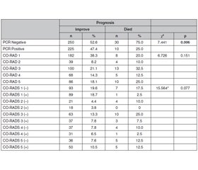
Three hundred six were male and 169 were female of the 515 patients and the mean age was 57.22 ± 19.77. According to the CO-RADS classification, 190 of the patients were CO-RADS 1 (36.9%), 43 of them were CO-RADS 2 (8.3%), 113 of them were CO-RADS 3 (21.9%), 73 of them were CO-RADS 4 (14.2%) and 96 of them were CO-RADS 5 (18.6%). Two hundred eighty patients (54.4%) had negative RT-PCR tests and 235 patients had positive RT-PCR tests. The comorbid disease rates were 40.4% HT, 16.7% DM, 13% asthma and/or COPD, 28% CAD, 12.6% psychiatric diseases, 3.7% oncologic diseases and 18.4% the other diseases. While 40 patients (7.8%) died, 475 patients (92.2%) recovered. Most of the patients who died were found to have a negative PCR test result (30 patients, 75%) and this difference was significant in the statistical analysis based on RT-PCR status (p = 0.006). 
A comparison of prognosis regarding the test results was shown in table 1. Moreover, evaluating the effects of comorbid diseases on prognosis, it was found that HT (p = 0.001), CAD (p = 0.004), and asthma and/or COPD (p = 0.019) were associated with poor prognosis. Psychiatric diseases (p = 0.143), DM (p = 0.143) and oncological diseases (p = 0.647) were not statistically significantly associated with prognosis. 
While no statistically significant difference was found between gender and prognosis (p = 0.478), age affected the prognosis significantly (Z = 6.448, p < 0.001). Improved: 57, interquartile range (IQR): 32 (years); died: 79, IQR: 15,50 (years). Comparison of prognosis concerning sex and comorbidities are shown in table 2. 
Additionally, it was seen that only age caused a significant difference in the model created with variables (categorical ones hypertension absent: 0, present: 1, Asthma-COPD absent: 0, present: 1, CAD absent: 0, present: 1, and age numerically) found to be effective on prognosis in paired comparisons (table 3).

Discussion

RT-PCR has been used in the diagnosis of COVID-19 since the beginning of the pandemic. However; low viral load, a mutant virus, inadequate swab samples, problems in transportation and storage of the material, and various factors related to the test kit affect the RT-PCR result. Although positive results can be obtained in some of the RT-PCR retests, they can not be obtained in any other patients. The false-negative rate in RT-PCR is reported as 1 and 30% [5, 6].
By evaluating the clinical conditions, laboratory tests, and chest CT findings of RT-PCR negative patients, COVID-19 can be diagnosed and their treatments can be planned. Some studies are reporting that chest CT is more sensitive than RT-PCR, especially in the early stage of the disease [7, 8].
CO-RADS offers a standardized assessment system that simplifies reporting with a five-point scale of suspected COVID-19 pulmonary involvement on chest CT [3]. Fu et al. examined the chest CT findings of two consecutive RT-PCR negative patients with suspected COVID-19 and emphasized that RT-PCR alone would not be sufficient to rule out the disease, and emphasized the importance of evaluating chest CT and RT-PCR together [9]. Chen et al. reported that early chest CT findings may have prognostic significance in RT-PCR negative patients [10]. It has also been reported that the less pulmonary consolidation found on chest CT, the higher probability of initial negative RT-PCR results [11]. Besides the chest CT findings in the RT-PCR negative group, the patient's clinical status and blood values were also taken into consideration in our study.
Various comorbid diseases are known to affect the prognosis of COVID-19. It has been reported that 10 to 34% of COVID-19 patients have HT [12]. Meng et al. reported in their meta-analysis that HT, DM, and cardiovascular diseases are important risk factors for the development of severe COVID-19 [13]. However, some studies have reported that HT is not associated with the development and prognosis of COVID-19. Kreutz et al. reported that although HT seems to be associated with more severe disease, higher risk of acute respiratory distress syndrome, and increased mortality, there is no strong evidence showing increased sensitivity of patients with HT [14]. Although it cannot be said clearly that HT affects the prognosis negatively, HT in old ages and different comorbidities with HT can negatively affect the prognosis. Cardiovascular diseases can lead to severe COVID-19, and also COVID-19 can cause heart damage [15].
There are several studies investigating the relation between DM and COVID-19. It has been reported that the rapid normalization of hyperglycemia during COVID-19 may contribute to a better prognosis by reducing the release of inflammatory cytokines and reducing the ACE2 binding capacity of the virus [16]. It has been reported that the presence of type 2 DM in intubated COVID-19 patients prolongs the healing process [17]. Considering that DM and HT increase body stress, cause vascular damage, and weaken immunity, it is obvious that they may play a role in the prognosis of COVID-19. Li et al. reported that patients with advanced age, hypertension, and high lactate dehydrogenase levels would need detailed observation and early intervention to prevent the potential development of severe COVID-19 [18]. The relationship of COVID-19 with coagulopathy has been demonstrated, and its association with vascular damage may worsen the prognosis of COVID-19. In our study, HT was found to be associated with poor prognosis, but no such relationship was found with DM. The stability of glucose regulation in DM patients included in our study may affect this result.
It can be said that patients with chronic respiratory diseases such as asthma and COPD have a low resistance to all respiratory viruses due to existing lung damage. In a study, including 44 672 confirmed COVID-19 patients, it was reported that chronic respiratory diseases were ranked 3rd in the mortality rate [19]. Based on this, it can be thought that controlling chronic respiratory diseases can reduce mortality and morbidity in COVID-19. In our study, similar to the literature, asthma and COPD were associated with poor prognosis.
It has been reported that the prognosis of COVID-19 is worse in patients with malignancy in some studies examining the relationship between various malignancies and COVID-19 [20, 21]. A total of 2% fatality rate was observed among the COVID-19 cases who had malignancies [22]. In patients with malignancy, the prognosis is expected to be poor because of chemotherapy, radiotherapy, and suppression of the immune system. N.M. Kuderer et al. reported that cancer patients are at high risk of mortality from COVID-19, regardless of whether they have active cancer, are receiving anticancer treatment, or both [20]. However, it should be kept in mind that conditions such as the type of malignancy, being in remission period, and having concurrent malignancies may have different effects on the prognosis. In our study, the prognosis relationship between COVID-19 and malignancies could not be determined because the number of patients with malignancy was low and the subtypes of malignancy were not determined.
There have been many studies on the effects of patients’ age on COVID-19 prognosis. It has been reported that while mortality rates due to COVID-19 are extremely low between the ages of 5 and 9, this rate increases linearly over the age of 30, and reaches the highest rate in patients over the age of 65 [23]. The hospitalization time has a positive correlation with advanced age. In addition, the high rate of comorbid diseases in advanced age also contributes to poor prognosis. In our study, the mortality rate was found high in elderly patients, similar to the literature.
In many studies, it has been reported that COVID-19 is more common in men. Y. Zhao et al. reported that ACE2 expression is dominant in Asian men, which may explain the higher incidence of COVID-19 in men [24]. It’s suggested that the high rate of smoking in men may explain this dominance. Similar to the literature, it was found that COVID-19 was more common in males, but no significant relationship was found between prognosis and gender in our study.
Mortality rates in RT-PCR negative patients who received COVID-19 treatment were higher than in those who were positive for RT-PCR in our study. Despite mild clinical conditions at the beginning of the disease, less mechanical ventilation, and the need for hospitalization in the intensive care unit in negative RT-PCR patients, it is reported that the mortality and hospitalization length are not different from RT-PCR positive patients [25]. The 30-day death rate of cancer patients with or without COVID-19 is poor, but the majority of deaths occur in RT-PCR-negative patients [26]. It has also been reported that patients with significant inflammatory responses in laboratory tests may have negative RT-PCR initially [27]. In some patients, the presence of the disease in the lower respiratory tract may cause negative RT-PCR, which may be associated with severe COVID-19. This situation may explain the high mortality rate in patients.RT-PCR positivity cannot be detected in some patients despite repeated RT-PCR tests, which may cause delays in treatment. It may be another reason for the high mortality rate in RT-PCR negative patients in our study. Serological tests may be recommended for this group of patients.
The limitations of our study are that the smoking status of the patients was not determined, the patients with more than one comorbidity were not evaluated as a different group and the mutant virus was not detected in groups. In addition, the small number of malignancy patients and the lack of evaluation according to the subtypes of malignancies may be other limitations of our study.

Conclusions

Even if RT-PCR is negative in the diagnosis of COVID-19 disease, RT-PCR should be repeated and a treatment plan should be applied by evaluating the hemogram values, chest CT findings, and the clinical condition of the patient.
Comorbidities of patients should be considered when planning treatment for COVID-19. Prospective studies in which patients with multiple comorbid diseases will be evaluated separately may support our study results. 
As the studies on RT-PCR negative COVID-19 patients increase, the reasons for the high mortality rate in these patients may be revealed.
 
Received 02.08.2022
Revised 11.09.2022
Accepted 21.09.2022

Список литературы

  1. Kim H., Hong H., Yoon S.H. Diagnostic performance of CT and reverse transcriptase polymerase chain reaction for coronavirus disease 2019: A meta-analysis. Radiology. 2020. 296(3). 145-55. https://doi.org/10.1148/radiol.2020201343.
  2. Li Y., Yao L., Li J., Chen L., Song Y., Cai Z. et al. Stability issues of RT-PCR testing of SARS-CoV-2 for hospitalized patients clinically diagnosed with COVID-19. J. Med. Virol. 2020. 92(7). 903-8. https://doi.org/10.1002/jmv.25786.
  3. Prokop M., vanEverdingen W., vanReesVellinga T., Quarles van Ufford H., Stöger L., Beenen L. et al. CO-RADS: A categorical CT assessment scheme for patients suspected of having COVID-19-Definition and evaluation. Radiology. 2020. 296(2). 97-104. https://doi.org/10.1148/radiol.2020201473.
  4. Guzik T.J., Mohiddin S.A., Dimarco A., Patel V., Savvatis K., Marelli-Berg F.M. et al. COVID-19 and the cardiovascular system: Implications for risk assessment, diagnosis, and treatment options. Cardiovasc. Res. 2020. 116(10). 1666-87. https://doi.org/10.1093/cvr/cvaa106.
  5. Long D.R., Gombar S., Hogan C.A., Greninger A.L., OReilly Shah V., Bryson-Cahn C. et al. Occurrence and timing of subsequent SARS-CoV-2 RT-PCR positivity among initially negative patients. MedRxiv2020:2020.05.03.20089151. https://doi.org/doi: 10.1101/2020.05.03.20089151.
  6. Arevalo-Rodriguez I., Buitrago-Garcia D., Simancas-Racines D., Zambrano-Achig P., Del Campo R., Ciapponi A. et al. False-negative results of initial RT-PCR assays for COVID-19: A systematic review. PLoS One. 2020. 15(12). e0242958. https://doi.org/10.1371/journal.pone.0242958.
  7. Khatami F., Saatchi M., Zadeh S.S.T., Aghamir Z.S., Shabestari A.N., Reis L.O. et al. A meta-analysis of accuracy and sensitivity of chest CT and RT-PCR in COVID-19 diagnosis. Sci. Rep. 2020. 10(1). 22402. https://doi.org/10.1038/s41598-020-80061-2.
  8. Fang Y., Zhang H., Xie J., Lin M., Ying L., Pang P. et al. Sensitivity of chest CT for COVID-19: Comparison to RT-PCR. Radiology. 2020. 296(2). 115-7. https://doi.org/10.1148/radiol.2020200432. 
  9. Fu Z., Tang N., Chen Y., Ma L., Wei Y., Lu Y. et al. CT features of COVID-19 patients with two consecutive negative RT-PCR tests after treatment. Sci. Rep. 2020. 10(1). 11548. https://doi.org/ 10.1038/s41598-020-68509-x. 
  10. Chen H.J., Qiu J., Wu B., Huang T., Gao Y., Wang Z.P. et al. Early chest CT features of patients with 2019 novel coronavirus (COVID-19) pneumonia: relationship to diagnosis and prognosis. Eur. Radiol. 2020. 30(11). 6178-85. https://doi.org/10.1007/s00330-020-06978-4.
  11. Chen D., Jiang X., Hong Y., Wen Z., Wei S., Peng G. et al. Can Chest CT Features distinguish patients with negative from those with positive initial RT-PCR results for Coronavirus Disease (COVID-19)? Am. J. Roentgenol. 2021. 216(1). 66-70. https://doi.org/10.2214/AJR.20.23012. 
  12. Tadic M., Cuspidi C., Grassi G., Mancia G. COVID-19 and arterial hypertension: Hypothesis or evidence? J. Clin Hypertens (Greenwich). 2020. 22(7). 1120-6. https://doi.org/10.1111/jch.13925. 
  13. Meng M., Zhao Q., Kumar R., Bai C., Deng Y., Wan B. Impact of cardiovascular and metabolic diseases on the severity of COVID-19: A systematic review and meta-analysis. Aging (Albany NY). 2020. 12(22). 23409-234. https://doi.org/10.18632/aging.
  14. Kreutz R., Algharably E., Azizi M., Dobrowolski P., Guzik T., Januszewicz A. et al. Hypertension, the renin-angiotensin system, and the risk of lower respiratory tract infections and lung injury: implications for COVID-19. Cardiovasc. Res. 2020. 116(10). 1688-99. https://doi.org/10.1093/cvr/cvaa097.
  15. Li B., Yang J., Zhao F., Zhi L., Wang X., Liu L. et al. Prevalence and impact of cardiovascular metabolic diseases on COVID-19 in China. Clin. Res. Cardiol. 2020. 109(5). 531-8. https://doi.org/ 10.1007/s00392-020-01626-9.
  16. Ceriello A. Hyperglycemia and the worse prognosis of COVID-19. Why a fast blood glucose control should be mandatory. Diabetes Res. Clin. Pract. 2020. 163. 108186. https://doi.org/10.1016/j.diabres.2020.108186. 
  17. Buetti N., Trimboli P., Mazzuchelli T., Lo Priore E., Balmelli C., Trkola A. et al. Diabetes mellitus is a risk factor for prolonged SARS-CoV-2 viral shedding in lower respiratory tract samples of critically ill patients. Endocrine. 2020. 70(3). 454-60. https://doi.org/10.1007/s12020-020-02465-4.
  18. Li X., Xu S., Yu M., Wang K., Tao Y., Zhou Y. et al. Risk factors for severity and mortality in adult COVID-19 inpatients in Wuhan. J. Allergy Clin. Immunol. 2020. 146(1). 110-8. https://doi.org/10.1016/j.jaci.2020.04.006.
  19. Novel Coronavirus Pneumonia Emergency Response Epidemiology T. The epidemiological characteristics of an outbreak of 2019 novel coronavirus diseases (COVID-19) in China. Zhonghua Liu Xing Bing Xue Za Zhi. 2020. 41(2). 145-51.
  20. Kuderer N.M., Choueiri T.K., Shah DP., Shyr Y., Rubinstein S.M., Rivera D.R. et al. Clinical impact of COVID-19 on patients with cancer (CCC19): A cohort study. Lancet. 2020. 395(10241). 1907-18. https://doi.org/10.1016/S0140-6736(20)31187-9.
  21. Yang K., Sheng Y., Huang C., Jin Y., Xiong N., Jiang K. et al. Clinical characteristics, outcomes, and risk factors for mortality in patients with cancer and COVID-19 in Hubei, China: a multicentre, retrospective, cohort study. Lancet Oncol. 2020. 21(7). 904-13. https://doi.org/10.1016/S1470-2045(20)30310-7.
  22. Wang D., Hu B., Hu C., Zhu F., Liu X., Zhang J. et al. Clinical characteristics of 138 hospitalized patients with 2019 novel coronavirus-infected pneumonia in Wuhan, China. JAMA. 2020. 323(11). 1061-9. https://doi.org/10.1001/jama.2020.1585.
  23. O’Driscoll M., Ribeiro Dos Santos G., Wang L., Cummings D.A.T., Azman A.S., Paireau J. et al. Age-specific mortality and immunity patterns of SARS-CoV-2. Nature. 2021. 590(7844). 140-5. https://doi.org/10.1038/s41586-020-2918-0.
  24. Zhao Y., Zhao Z., Wang Y., Zhou Y., Ma Y., Zuo W. Single-cell RNA expression profiling of ACE2, the putative receptor of Wuhan 2019-nCov. BioRxiv. 2020.01.26.919985. https://doi.org/ 10.1101/2020.01.26.919985.
  25. Baicry F., Le Borgne P., Fabacher T., Behr M., Lemaitre E.L., Gayol PA. et al. Patients with initial negative RT-PCR and typical imaging of COVID-19: Clinical implications. J. Clin. Med. 2020. 9(9). 3014. https://doi.org/10.3390/jcm9093014.
  26. Assaad S., Avrillon V., Fournier M.L, Mastroianni B., Russias B., Swalduz A. et al. High mortality rate in cancer patients with symptoms of COVID-19 with or without detectable SARS-COV-2 on RT-PCR. Eur. J. Cancer. 2020. 135. 251-9. https://doi.org/10.1016/ j.ejca.2020.05.028.
  27. Lascarrou J.B., Colin G., Le Thuaut A., Serck N., Ohana M., Sauneuf B. et al. Predictors of negative first SARS-CoV-2 RT-PCR despite final diagnosis of COVID-19 and association with outcome. Sci. Rep. 2021. 11(1). 2388. https://doi.org/10.1038/s41598-021-82192-6.

Вернуться к номеру